Crystal structure of N-domain of FKBP22 from Shewanella sp. SIB1: dimer dissociation by disruption of Val-Leu knot
Budiman, C., Angkawidjaja, C., Motoike, H., Koga, Y., Takano, K., Kanaya, S.(2011) Protein Sci 20: 1755-1764
- PubMed: 21837652 Search on PubMedSearch on PubMed Central
- DOI: https://doi.org/10.1002/pro.714
- Primary Citation Related Structures:
3B09 - PubMed Abstract:
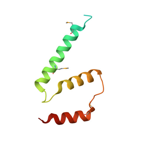
FK506-binding protein 22 (FKBP22) from the psychrotophic bacterium Shewanella sp. SIB1 (SIB1 FKBP22) is a homodimeric protein with peptidyl prolyl cis-trans isomerase (PPIase) activity. Each monomer consists of the N-terminal domain responsible for dimerization and C-terminal catalytic domain. To reveal interactions at the dimer interface of SIB1 FKBP22, the crystal structure of the N-domain of SIB1 FKBP22 (SN-FKBP22, residues 1-68) was determined at 1.9 Å resolution. SN-FKBP22 forms a dimer, in which each monomer consists of three helices (α1, α2, and α3N). In the dimer, two monomers have head-to-head interactions, in which residues 8-64 of one monomer form tight interface with the corresponding residues of the other. The interface is featured by the presence of a Val-Leu knot, in which Val37 and Leu41 of one monomer interact with Val41 and Leu37 of the other, respectively. To examine whether SIB1 FKBP22 is dissociated into the monomers by disruption of this knot, the mutant protein V37R/L41R-FKBP22, in which Val37 and Leu41 of SIB1 FKBP22 are simultaneously replaced by Arg, was constructed and biochemically characterized. This mutant protein was indistinguishable from the SIB1 FKBP22 derivative lacking the N-domain in oligomeric state, far-UV CD spectrum, thermal denaturation curve, PPIase activity, and binding ability to a folding intermediate of protein, suggesting that the N-domain of V37R/L41R-FKBP22 is disordered. We propose that a Val-Leu knot at the dimer interface of SIB1 FKBP22 is important for dimerization and dimerization is required for folding of the N-domain.
- Department of Material and Life Science, Graduate School of Engineering, Osaka University, Suita, Osaka 565-0871, Japan.
Organizational Affiliation: