3AXZ | pdb_00003axz

Crystal structure of Haemophilus influenzae TrmD in complex with adenosine


Experimental Data Snapshot

  • Method: X-RAY DIFFRACTION
  • Resolution: 2.25 Å
  • R-Value Free: 
    0.243 (Depositor), 0.243 (DCC) 
  • R-Value Work: 
    0.189 (Depositor), 0.189 (DCC) 

Starting Model: experimental
View more details

wwPDB Validation 3D Report Full Report

Validation slider image for 3AXZ

Ligand Structure Quality Assessment 


This is version 1.1 of the entry. See complete history

Literature

Differentiating analogous tRNA methyltransferases by fragments of the methyl donor.

Lahoud, G.Goto-Ito, S.Yoshida, K.Ito, T.Yokoyama, S.Hou, Y.M.

(2011) RNA 17: 1236-1246

  • DOI: https://doi.org/10.1261/rna.2706011
  • Primary Citation Related Structures: 
    3AXZ, 3AY0

  • PubMed Abstract: 

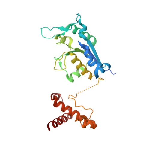
    Bacterial TrmD and eukaryotic-archaeal Trm5 form a pair of analogous tRNA methyltransferase that catalyze methyl transfer from S-adenosyl methionine (AdoMet) to N(1) of G37, using catalytic motifs that share no sequence or structural homology. Here we show that natural and synthetic analogs of AdoMet are unable to distinguish TrmD from Trm5. Instead, fragments of AdoMet, adenosine and methionine, are selectively inhibitory of TrmD rather than Trm5. Detailed structural information of the two enzymes in complex with adenosine reveals how Trm5 escapes targeting by adopting an altered structure, whereas TrmD is trapped by targeting due to its rigid structure that stably accommodates the fragment. Free energy analysis exposes energetic disparities between the two enzymes in how they approach the binding of AdoMet versus fragments and provides insights into the design of inhibitors selective for TrmD.


  • Organizational Affiliation
    • Thomas Jefferson University, Department of Biochemistry and Molecular Biology, Philadelphia, Pennsylvania 19107, USA.

Macromolecule Content 

  • Total Structure Weight: 30.02 kDa 
  • Atom Count: 2,040 
  • Modeled Residue Count: 241 
  • Deposited Residue Count: 266 
  • Unique protein chains: 1

Macromolecules

Find similar proteins by:|  3D Structure
Entity ID: 1
MoleculeChains  Sequence LengthOrganismDetailsImage
tRNA (guanine-N(1)-)-methyltransferase266Haemophilus influenzae Rd KW20Mutation(s): 0 
Gene Names: HI_0202trmD
EC: 2.1.1.31 (PDB Primary Data), 2.1.1.228 (UniProt)
UniProt
Find proteins for P43912 (Haemophilus influenzae (strain ATCC 51907 / DSM 11121 / KW20 / Rd))
Explore P43912 
Go to UniProtKB:  P43912
Entity Groups
Sequence Clusters30% Identity50% Identity70% Identity90% Identity95% Identity100% Identity
UniProt GroupP43912
Sequence Annotations
Expand
Reference Sequence

Small Molecules

Ligands 1 Unique
IDChains Name / Formula / InChI Key2D Diagram3D Interactions
ADN

Query on ADN


Download:Ideal Coordinates CCD File
B [auth A]ADENOSINE
C10 H13 N5 O4
OIRDTQYFTABQOQ-KQYNXXCUSA-N

Experimental Data & Validation

Experimental Data

  • Method: X-RAY DIFFRACTION
  • Resolution: 2.25 Å
  • R-Value Free:  0.243 (Depositor), 0.243 (DCC) 
  • R-Value Work:  0.189 (Depositor), 0.189 (DCC) 
Space Group: H 3 2
Unit Cell:
Length ( Å )Angle ( ˚ )
a = 94.97α = 90
b = 94.97β = 90
c = 177.89γ = 120
Software Package:
Software NamePurpose
HKL-2000data collection
PHASERphasing
CNSrefinement
HKL-2000data reduction
HKL-2000data scaling

Structure Validation

View Full Validation Report


Ligand Structure Quality Assessment 


Entry History 

Deposition Data

Revision History  (Full details and data files)

  • Version 1.0: 2011-08-17
    Type: Initial release
  • Version 1.1: 2023-11-01
    Changes: Data collection, Database references, Derived calculations, Refinement description