3AUN

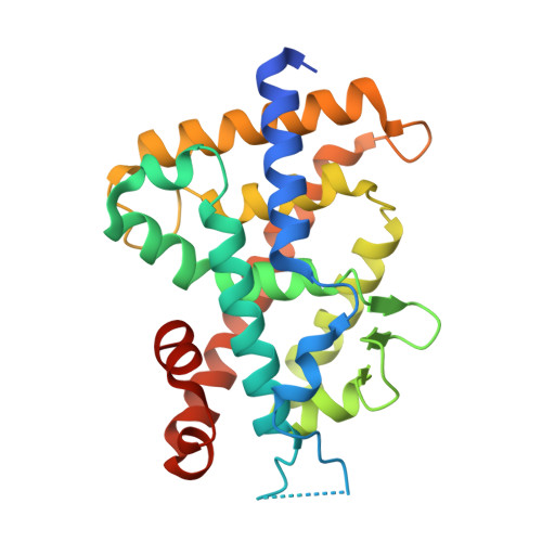
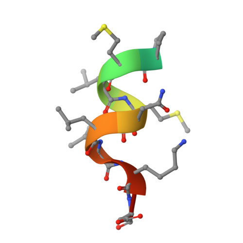
Crystal structure of the rat vitamin D receptor ligand binding domain complexed with YR335 and a synthetic peptide containing the NR2 box of DRIP 205


Experimental Data Snapshot

  • Method: X-RAY DIFFRACTION
  • Resolution: 1.81 Å
  • R-Value Free: 0.237 
  • R-Value Work: 0.196 
  • R-Value Observed: 0.198 

Starting Model: experimental
View more details

wwPDB Validation   3D Report Full Report


Ligand Structure Quality Assessment 


This is version 1.4 of the entry. See complete history


Literature

Design, synthesis and X-ray crystallographic study of new nonsecosteroidal vitamin D receptor ligands

Demizu, Y.Takahashi, T.Kaneko, F.Sato, Y.Okuda, H.Ochiai, E.Horie, K.Takagi, K.Kakuda, S.Takimoto-Kamimura, M.Kurihara, M.

(2011) Bioorg Med Chem Lett 21: 6104-6107

  • DOI: https://doi.org/10.1016/j.bmcl.2011.08.047
  • Primary Citation of Related Structures:  
    3AUN

  • PubMed Abstract: 

    We designed and synthesized nonsecosteroidal vitamin D receptor (VDR) ligands that formed H-bonds with six amino acid residues (Tyr143, Ser233, Arg270, Ser274, His301 and His393) of the VDR ligand-binding domain. The ligand YR335 exhibited potent transcriptional activity, which was comparable to those of 1α,25-dihydroxyvitamin D(3) and YR301. The crystal structure of the complex formed between YR335 and the VDR ligand-binding domain was solved, which revealed that YR335 formed H-bonds with the six amino acid residues mentioned above.


  • Organizational Affiliation

    Division of Organic Chemistry, National Institute of Health Sciences, 1-18-1, Kamiyoga, Setagaya, Tokyo 158-8501, Japan.


Macromolecules
Find similar proteins by:  (by identity cutoff)  |  3D Structure
Entity ID: 1
MoleculeChains Sequence LengthOrganismDetailsImage
Vitamin D3 receptor265Rattus norvegicusMutation(s): 0 
UniProt
Find proteins for P13053 (Rattus norvegicus)
Explore P13053 
Go to UniProtKB:  P13053
Entity Groups  
Sequence Clusters30% Identity50% Identity70% Identity90% Identity95% Identity100% Identity
UniProt GroupP13053
Sequence Annotations
Expand
  • Reference Sequence

Find similar proteins by:  Sequence   |   3D Structure  

Entity ID: 2
MoleculeChains Sequence LengthOrganismDetailsImage
DRIP 205 NR2 box peptide13synthetic constructMutation(s): 0 
UniProt & NIH Common Fund Data Resources
Find proteins for Q15648 (Homo sapiens)
Explore Q15648 
Go to UniProtKB:  Q15648
PHAROS:  Q15648
GTEx:  ENSG00000125686 
Entity Groups  
Sequence Clusters30% Identity50% Identity70% Identity90% Identity95% Identity100% Identity
UniProt GroupQ15648
Sequence Annotations
Expand
  • Reference Sequence
Small Molecules
Ligands 1 Unique
IDChains Name / Formula / InChI Key2D Diagram3D Interactions
YR4
Query on YR4

Download Ideal Coordinates CCD File 
C [auth A](2R)-2-{4-[3-(4-{[(2R)-2-hydroxy-3,3-dimethylbutyl]oxy}-3-methylphenyl)pentan-3-yl]-2-methylphenoxy}butane-1,4-diol
C29 H44 O5
DKNLNQKPISQAEP-SQHAQQRYSA-N
Experimental Data & Validation

Experimental Data

  • Method: X-RAY DIFFRACTION
  • Resolution: 1.81 Å
  • R-Value Free: 0.237 
  • R-Value Work: 0.196 
  • R-Value Observed: 0.198 
  • Space Group: P 21 21 2
Unit Cell:
Length ( Å )Angle ( ˚ )
a = 44.237α = 90
b = 136.521β = 90
c = 47.334γ = 90
Software Package:
Software NamePurpose
HKL-2000data collection
MOLREPphasing
REFMACrefinement
HKL-2000data reduction
HKL-2000data scaling

Structure Validation

View Full Validation Report


Ligand Structure Quality Assessment 


Entry History 

Deposition Data

Revision History  (Full details and data files)

  • Version 1.0: 2012-02-15
    Type: Initial release
  • Version 1.1: 2012-08-01
    Changes: Database references
  • Version 1.2: 2017-08-16
    Changes: Refinement description, Source and taxonomy
  • Version 1.3: 2017-08-23
    Changes: Source and taxonomy
  • Version 1.4: 2023-11-01
    Changes: Data collection, Database references, Derived calculations, Refinement description