3AUA

Crystal structure of the quaternary complex-2 of an isomerase


Experimental Data Snapshot

  • Method: X-RAY DIFFRACTION
  • Resolution: 2.15 Å
  • R-Value Free: 0.220 
  • R-Value Work: 0.172 
  • R-Value Observed: 0.175 

wwPDB Validation   3D Report Full Report


Ligand Structure Quality Assessment 


This is version 1.2 of the entry. See complete history


Literature

Molecular basis of fosmidomycin's action on the human malaria parasite Plasmodium falciparum

Umeda, T.Tanaka, N.Kusakabe, Y.Nakanishi, M.Kitade, Y.Nakamura, K.T.

(2011) Sci Rep 1: 9-9

  • DOI: https://doi.org/10.1038/srep00009
  • Primary Citation of Related Structures:  
    3AU8, 3AU9, 3AUA

  • PubMed Abstract: 

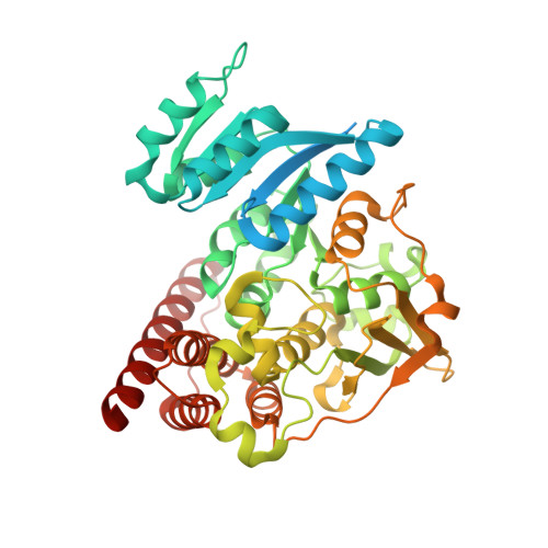
    The human malaria parasite Plasmodium falciparum is responsible for the deaths of more than a million people each year. Fosmidomycin has been proven to be efficient in the treatment of P. falciparum malaria by inhibiting 1-deoxy-D-xylulose 5-phosphate reductoisomerase (DXR), an enzyme of the non-mevalonate pathway, which is absent in humans. However, the structural details of DXR inhibition by fosmidomycin in P. falciparum are unknown. Here, we report the crystal structures of fosmidomycin-bound complete quaternary complexes of PfDXR. Our study revealed that (i) an intrinsic flexibility of the PfDXR molecule accounts for an induced-fit movement to accommodate the bound inhibitor in the active site and (ii) a cis arrangement of the oxygen atoms of the hydroxamate group of the bound inhibitor is essential for tight binding of the inhibitor to the active site metal. We expect the present structures to be useful guides for the design of more effective antimalarial compounds.


  • Organizational Affiliation

    School of Pharmacy, Showa University, Tokyo 142-8555, Japan.


Macromolecules
Find similar proteins by:  (by identity cutoff)  |  3D Structure
Entity ID: 1
MoleculeChains Sequence LengthOrganismDetailsImage
1-deoxy-D-xylulose 5-phosphate reductoisomerase
A, B
488Plasmodium falciparumMutation(s): 0 
Gene Names: dxr
UniProt
Find proteins for O96693 (Plasmodium falciparum (isolate HB3))
Explore O96693 
Go to UniProtKB:  O96693
Entity Groups  
Sequence Clusters30% Identity50% Identity70% Identity90% Identity95% Identity100% Identity
UniProt GroupO96693
Sequence Annotations
Expand
  • Reference Sequence
Small Molecules
Ligands 4 Unique
IDChains Name / Formula / InChI Key2D Diagram3D Interactions
NDP
Query on NDP

Download Ideal Coordinates CCD File 
C [auth A],
F [auth B]
NADPH DIHYDRO-NICOTINAMIDE-ADENINE-DINUCLEOTIDE PHOSPHATE
C21 H30 N7 O17 P3
ACFIXJIJDZMPPO-NNYOXOHSSA-N
F98
Query on F98

Download Ideal Coordinates CCD File 
E [auth A],
H [auth B]
3-[ethanoyl(hydroxy)amino]propylphosphonic acid
C5 H12 N O5 P
PKMNDDZSIHLLLI-UHFFFAOYSA-N
CA
Query on CA

Download Ideal Coordinates CCD File 
I [auth B],
J [auth B]
CALCIUM ION
Ca
BHPQYMZQTOCNFJ-UHFFFAOYSA-N
MG
Query on MG

Download Ideal Coordinates CCD File 
D [auth A],
G [auth B]
MAGNESIUM ION
Mg
JLVVSXFLKOJNIY-UHFFFAOYSA-N
Experimental Data & Validation

Experimental Data

  • Method: X-RAY DIFFRACTION
  • Resolution: 2.15 Å
  • R-Value Free: 0.220 
  • R-Value Work: 0.172 
  • R-Value Observed: 0.175 
  • Space Group: P 1 21 1
Unit Cell:
Length ( Å )Angle ( ˚ )
a = 51.011α = 90
b = 77.606β = 90.63
c = 111.736γ = 90
Software Package:
Software NamePurpose
ADSCdata collection
REFMACrefinement
HKL-2000data reduction
HKL-2000data scaling
REFMACphasing

Structure Validation

View Full Validation Report


Ligand Structure Quality Assessment 


Entry History 

Deposition Data

Revision History  (Full details and data files)

  • Version 1.0: 2011-08-10
    Type: Initial release
  • Version 1.1: 2013-06-12
    Changes: Database references
  • Version 1.2: 2024-03-13
    Changes: Data collection, Database references, Derived calculations