3ASL | pdb_00003asl

Structure of UHRF1 in complex with histone tail


Experimental Data Snapshot

  • Method: X-RAY DIFFRACTION
  • Resolution: 1.41 Å
  • R-Value Free: 
    0.196 (Depositor), 0.268 (DCC) 
  • R-Value Work: 
    0.159 (Depositor), 0.236 (DCC) 
  • R-Value Observed: 
    0.161 (Depositor) 

wwPDB Validation 3D Report Full Report

Validation slider image for 3ASL

This is version 1.3 of the entry. See complete history

Literature

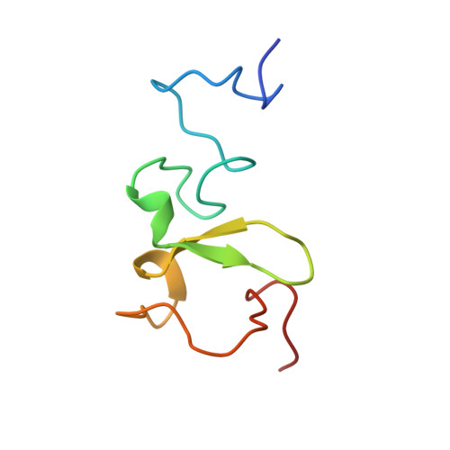
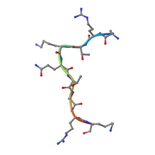
Recognition of modification status on a histone H3 tail by linked histone reader modules of the epigenetic regulator UHRF1

Arita, K.Isogai, S.Oda, T.Unoki, M.Sugita, K.Sekiyama, N.Kuwata, K.Hamamoto, R.Tochio, H.Sato, M.Ariyoshi, M.Shirakawa, M.

(2012) Proc Natl Acad Sci U S A 109: 12950-12955

  • DOI: https://doi.org/10.1073/pnas.1203701109
  • Primary Citation Related Structures: 
    3ASK, 3ASL

  • PubMed Abstract: 

    Multiple covalent modifications on a histone tail are often recognized by linked histone reader modules. UHRF1 [ubiquitin-like, containing plant homeodomain (PHD) and really interesting new gene (RING) finger domains 1], an essential factor for maintenance of DNA methylation, contains linked two-histone reader modules, a tandem Tudor domain and a PHD finger, tethered by a 17-aa linker, and has been implicated to link histone modifications and DNA methylation. Here, we present the crystal structure of the linked histone reader modules of UHRF1 in complex with the amino-terminal tail of histone H3. Our structural and biochemical data provide the basis for combinatorial readout of unmodified Arg-2 (H3-R2) and methylated Lys-9 (H3-K9) by the tandem tudor domain and the PHD finger. The structure reveals that the intermodule linker plays an essential role in the formation of a histone H3-binding hole between the reader modules by making extended contacts with the tandem tudor domain. The histone H3 tail fits into the hole by adopting a compact fold harboring a central helix, which allows both of the reader modules to simultaneously recognize the modification states at H3-R2 and H3-K9. Our data also suggest that phosphorylation of a linker residue can modulate the relative position of the reader modules, thereby altering the histone H3-binding mode. This finding implies that the linker region plays a role as a functional switch of UHRF1 involved in multiple regulatory pathways such as maintenance of DNA methylation and transcriptional repression.


  • Organizational Affiliation
    • Department of Molecular Engineering, Graduate School of Engineering, Kyoto University, Kyoto 615-8510, Japan.

Macromolecule Content 

  • Total Structure Weight: 9.68 kDa 
  • Atom Count: 696 
  • Modeled Residue Count: 77 
  • Deposited Residue Count: 81 
  • Unique protein chains: 2

Macromolecules

Find similar proteins by:|  3D Structure
Entity ID: 1
MoleculeChains  Sequence LengthOrganismDetailsImage
E3 ubiquitin-protein ligase UHRF170Homo sapiensMutation(s): 0 
Gene Names: UHRF1
EC: 6.3.2 (PDB Primary Data), 2.3.2.27 (UniProt)
UniProt & NIH Common Fund Data Resources
Find proteins for Q96T88 (Homo sapiens)
Explore Q96T88 
Go to UniProtKB:  Q96T88
PHAROS:  Q96T88
GTEx:  ENSG00000276043 
Entity Groups
Sequence Clusters30% Identity50% Identity70% Identity90% Identity95% Identity100% Identity
UniProt GroupQ96T88
Sequence Annotations
Expand
Reference Sequence
Find similar proteins by:  Sequence   |   3D Structure  
Entity ID: 2
MoleculeChains  Sequence LengthOrganismDetailsImage
Histone H3.311Homo sapiensMutation(s): 0 
UniProt & NIH Common Fund Data Resources
Find proteins for P68431 (Homo sapiens)
Explore P68431 
Go to UniProtKB:  P68431
Entity Groups
Sequence Clusters30% Identity50% Identity70% Identity90% Identity95% Identity100% Identity
UniProt GroupP68431
Sequence Annotations
Expand
Reference Sequence

Small Molecules

Ligands 2 Unique
IDChains Name / Formula / InChI Key2D Diagram3D Interactions
ZN

Query on ZN


Download:Ideal Coordinates CCD File
C [auth A],
D [auth A],
E [auth A],
F [auth A]
ZINC ION
Zn
PTFCDOFLOPIGGS-UHFFFAOYSA-N
EDO

Query on EDO


Download:Ideal Coordinates CCD File
G [auth A],
H [auth A],
I [auth A],
J [auth B]
1,2-ETHANEDIOL
C2 H6 O2
LYCAIKOWRPUZTN-UHFFFAOYSA-N

Experimental Data & Validation

Experimental Data

  • Method: X-RAY DIFFRACTION
  • Resolution: 1.41 Å
  • R-Value Free:  0.196 (Depositor), 0.268 (DCC) 
  • R-Value Work:  0.159 (Depositor), 0.236 (DCC) 
  • R-Value Observed: 0.161 (Depositor) 
Space Group: C 1 2 1
Unit Cell:
Length ( Å )Angle ( ˚ )
a = 67.21α = 90
b = 36.137β = 110.69
c = 34.175γ = 90
Software Package:
Software NamePurpose
ADSCdata collection
SHELXSphasing
REFMACrefinement
HKL-2000data reduction
HKL-2000data scaling

Structure Validation

View Full Validation Report


Entry History 

Deposition Data

Revision History  (Full details and data files)

  • Version 1.0: 2012-01-25
    Type: Initial release
  • Version 1.1: 2012-08-15
    Changes: Database references
  • Version 1.2: 2013-06-05
    Changes: Database references
  • Version 1.3: 2024-03-13
    Changes: Data collection, Database references, Derived calculations