3AQL | pdb_00003aql

Structure of bacterial protein (apo form II)


Experimental Data Snapshot

  • Method: X-RAY DIFFRACTION
  • Resolution: 3.00 Å
  • R-Value Free: 
    0.251 (Depositor), 0.251 (DCC) 
  • R-Value Work: 
    0.237 (Depositor), 0.237 (DCC) 
  • R-Value Observed: 
    0.237 (Depositor) 

wwPDB Validation 3D Report Full Report

Validation slider image for 3AQL

This is version 1.3 of the entry. See complete history

Literature

Mechanism for the alteration of the substrate specificities of template-independent RNA polymerases

Toh, Y.Takeshita, D.Nagaike, T.Numata, T.Tomita, K.

(2011) Structure 19: 232-243

  • DOI: https://doi.org/10.1016/j.str.2010.12.006
  • Primary Citation Related Structures: 
    3AQK, 3AQL, 3AQM, 3AQN

  • PubMed Abstract: 

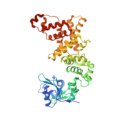
    PolyA polymerase (PAP) adds a polyA tail onto the 3'-end of RNAs without a nucleic acid template, using adenosine-5'-triphosphate (ATP) as a substrate. The mechanism for the substrate selection by eubacterial PAP remains obscure. Structural and biochemical studies of Escherichia coli PAP (EcPAP) revealed that the shape and size of the nucleobase-interacting pocket of EcPAP are maintained by an intra-molecular hydrogen-network, making it suitable for the accommodation of only ATP, using a single amino acid, Arg(197). The pocket structure is sustained by interactions between the catalytic domain and the RNA-binding domain. EcPAP has a flexible basic C-terminal region that contributes to optimal RNA translocation for processive adenosine 5'-monophosphate (AMP) incorporations onto the 3'-end of RNAs. A comparison of the EcPAP structure with those of other template-independent RNA polymerases suggests that structural changes of domain(s) outside the conserved catalytic core domain altered the substrate specificities of the template-independent RNA polymerases.


  • Organizational Affiliation
    • Biomedical Research Institute, National Institute of Advanced Industrial Science and Technology, 1-1-1, Higashi, Tsukuba-shi, Ibaraki 305-8566, Japan.

Macromolecule Content 

  • Total Structure Weight: 96.26 kDa 
  • Atom Count: 6,342 
  • Modeled Residue Count: 776 
  • Deposited Residue Count: 830 
  • Unique protein chains: 1

Macromolecules

Find similar proteins by:|  3D Structure
Entity ID: 1
MoleculeChains  Sequence LengthOrganismDetailsImage
Poly(A) polymerase
A, B
415Escherichia coli DH1Mutation(s): 1 
Gene Names: EcDH1_3459
EC: 2.7.7.19

Experimental Data & Validation

Experimental Data

  • Method: X-RAY DIFFRACTION
  • Resolution: 3.00 Å
  • R-Value Free:  0.251 (Depositor), 0.251 (DCC) 
  • R-Value Work:  0.237 (Depositor), 0.237 (DCC) 
  • R-Value Observed: 0.237 (Depositor) 
Space Group: P 42 21 2
Unit Cell:
Length ( Å )Angle ( ˚ )
a = 133.012α = 90
b = 133.012β = 90
c = 176.845γ = 90
Software Package:
Software NamePurpose
HKL-2000data collection
PHASERphasing
CNSrefinement
HKL-2000data reduction
HKL-2000data scaling

Structure Validation

View Full Validation Report


Entry History 

Deposition Data

Revision History  (Full details and data files)

  • Version 1.0: 2011-02-09
    Type: Initial release
  • Version 1.1: 2011-07-13
    Changes: Version format compliance
  • Version 1.2: 2013-07-31
    Changes: Database references
  • Version 1.3: 2024-03-13
    Changes: Data collection, Database references, Derived calculations