3APS | pdb_00003aps

Crystal structure of Trx4 domain of ERdj5


Experimental Data Snapshot

  • Method: X-RAY DIFFRACTION
  • Resolution: 1.90 Å
  • R-Value Free: 
    0.242 (Depositor), 0.238 (DCC) 
  • R-Value Work: 
    0.206 (Depositor), 0.203 (DCC) 
  • R-Value Observed: 
    0.208 (Depositor) 

wwPDB Validation 3D Report Full Report

Validation slider image for 3APS

This is version 1.2 of the entry. See complete history

Literature

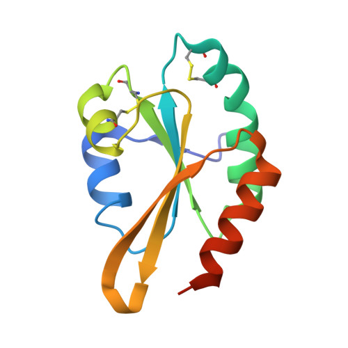
Structural basis of an ERAD pathway mediated by the ER-resident protein disulfide reductase ERdj5.

Hagiwara, M.Maegawa, K.Suzuki, M.Ushioda, R.Araki, K.Matsumoto, Y.Hoseki, J.Nagata, K.Inaba, K.

(2011) Mol Cell 41: 432-444

  • DOI: https://doi.org/10.1016/j.molcel.2011.01.021
  • Primary Citation Related Structures: 
    3APO, 3APQ, 3APS

  • PubMed Abstract: 

    ER-associated degradation (ERAD) is an ER quality-control process that eliminates terminally misfolded proteins. ERdj5 was recently discovered to be a key ER-resident PDI family member protein that accelerates ERAD by reducing incorrect disulfide bonds in misfolded glycoproteins recognized by EDEM1. We here solved the crystal structure of full-length ERdj5, thereby revealing that ERdj5 contains the N-terminal J domain and six tandem thioredoxin domains that can be divided into the N- and C-terminal clusters. Our systematic biochemical analyses indicated that two thioredoxin domains that constitute the C-terminal cluster form the highly reducing platform that interacts with EDEM1 and reduces EDEM1-recruited substrates, leading to their facilitated degradation. The pulse-chase experiment further provided direct evidence for the sequential movement of an ERAD substrate from calnexin to the downstream EDEM1-ERdj5 complex, and then to the retrotranslocation channel, probably through BiP. We present a detailed molecular view of how ERdj5 mediates ERAD in concert with EDEM1.


  • Organizational Affiliation
    • Department of Molecular and Cellular Biology, Institute for Frontier Medical Sciences, Kyoto University, Kyoto 606-8507, Japan.

Macromolecule Content 

  • Total Structure Weight: 28.07 kDa 
  • Atom Count: 1,915 
  • Modeled Residue Count: 228 
  • Deposited Residue Count: 244 
  • Unique protein chains: 1

Macromolecules

Find similar proteins by:|  3D Structure
Entity ID: 1
MoleculeChains  Sequence LengthOrganismDetailsImage
DnaJ homolog subfamily C member 10
A, B
122Mus musculusMutation(s): 0 
Gene Names: DNAJC10ERDJ5JPDI
EC: 1.8.4.2
UniProt & NIH Common Fund Data Resources
Find proteins for Q9DC23 (Mus musculus)
Explore Q9DC23 
Go to UniProtKB:  Q9DC23
IMPC:  MGI:1914111
Entity Groups
Sequence Clusters30% Identity50% Identity70% Identity90% Identity95% Identity100% Identity
UniProt GroupQ9DC23
Sequence Annotations
Expand
Reference Sequence

Experimental Data & Validation

Experimental Data

  • Method: X-RAY DIFFRACTION
  • Resolution: 1.90 Å
  • R-Value Free:  0.242 (Depositor), 0.238 (DCC) 
  • R-Value Work:  0.206 (Depositor), 0.203 (DCC) 
  • R-Value Observed: 0.208 (Depositor) 
Space Group: P 1 21 1
Unit Cell:
Length ( Å )Angle ( ˚ )
a = 56.06α = 90
b = 33.419β = 92.85
c = 68.343γ = 90
Software Package:
Software NamePurpose
HKL-2000data collection
SHELXmodel building
REFMACrefinement
HKL-2000data reduction
SCALEPACKdata scaling
SHELXphasing

Structure Validation

View Full Validation Report


Entry History 

Deposition Data

Revision History  (Full details and data files)

  • Version 1.0: 2011-04-20
    Type: Initial release
  • Version 1.1: 2011-07-13
    Changes: Version format compliance
  • Version 1.2: 2024-10-23
    Changes: Data collection, Database references, Derived calculations, Structure summary