3AHQ

hyperactive human Ero1


Experimental Data Snapshot

  • Method: X-RAY DIFFRACTION
  • Resolution: 2.35 Å
  • R-Value Free: 0.282 
  • R-Value Work: 0.237 
  • R-Value Observed: 0.239 

wwPDB Validation   3D Report Full Report


Ligand Structure Quality Assessment 


This is version 1.2 of the entry. See complete history


Literature

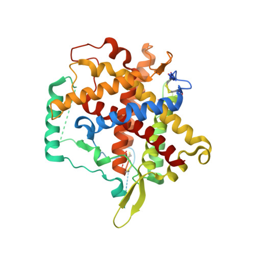
Crystal structures of human Ero1-alpha reveal the mechanisms of regulated and targeted oxidation of PDI

Inaba, K.Masui, S.Iida, H.Vavassori, S.Sitia, R.Suzuki, M.

(2010) EMBO J 29: 3330-3343

  • DOI: https://doi.org/10.1038/emboj.2010.222
  • Primary Citation of Related Structures:  
    3AHQ, 3AHR

  • PubMed Abstract: 

    In the endoplasmic reticulum (ER) of eukaryotic cells, Ero1 flavoenzymes promote oxidative protein folding through protein disulphide isomerase (PDI), generating reactive oxygen species (hydrogen peroxide) as byproducts. Therefore, Ero1 activity must be strictly regulated to avoid futile oxidation cycles in the ER. Although regulatory mechanisms restraining Ero1α activity ensure that not all PDIs are oxidized, its specificity towards PDI could allow other resident oxidoreductases to remain reduced and competent to carry out isomerization and reduction of protein substrates. In this study, crystal structures of human Ero1α were solved in its hyperactive and inactive forms. Our findings reveal that human Ero1α modulates its oxidative activity by properly positioning regulatory cysteines within an intrinsically flexible loop, and by fine-tuning the electron shuttle ability of the loop through disulphide rearrangements. Specific PDI targeting is guaranteed by electrostatic and hydrophobic interactions of Ero1α with the PDI b'-domain through its substrate-binding pocket. These results reveal the molecular basis of the regulation and specificity of protein disulphide formation in human cells.


  • Organizational Affiliation

    Division of Protein Chemistry, Post-Genome Science Center, Medical Institute of Bioregulation, Kyushu University, Maidashi, Higashi-ku, Fukuoka, Japan. inaba-k@bioreg.kyushu-u.ac.jp


Macromolecules
Find similar proteins by:  (by identity cutoff)  |  3D Structure
Entity ID: 1
MoleculeChains Sequence LengthOrganismDetailsImage
ERO1-like protein alpha465Homo sapiensMutation(s): 3 
Gene Names: Ero1-Lalpha
EC: 1.8.4
UniProt & NIH Common Fund Data Resources
Find proteins for Q96HE7 (Homo sapiens)
Explore Q96HE7 
Go to UniProtKB:  Q96HE7
PHAROS:  Q96HE7
GTEx:  ENSG00000197930 
Entity Groups  
Sequence Clusters30% Identity50% Identity70% Identity90% Identity95% Identity100% Identity
UniProt GroupQ96HE7
Sequence Annotations
Expand
  • Reference Sequence
Small Molecules
Ligands 1 Unique
IDChains Name / Formula / InChI Key2D Diagram3D Interactions
FAD
Query on FAD

Download Ideal Coordinates CCD File 
B [auth A]FLAVIN-ADENINE DINUCLEOTIDE
C27 H33 N9 O15 P2
VWWQXMAJTJZDQX-UYBVJOGSSA-N
Experimental Data & Validation

Experimental Data

  • Method: X-RAY DIFFRACTION
  • Resolution: 2.35 Å
  • R-Value Free: 0.282 
  • R-Value Work: 0.237 
  • R-Value Observed: 0.239 
  • Space Group: I 2 2 2
Unit Cell:
Length ( Å )Angle ( ˚ )
a = 54.962α = 90
b = 140.73β = 90
c = 144.855γ = 90
Software Package:
Software NamePurpose
BSSdata collection
MOLREPphasing
REFMACrefinement
HKL-2000data reduction
HKL-2000data scaling

Structure Validation

View Full Validation Report


Ligand Structure Quality Assessment 


Entry History 

Deposition Data

Revision History  (Full details and data files)

  • Version 1.0: 2010-12-22
    Type: Initial release
  • Version 1.1: 2011-07-13
    Changes: Version format compliance
  • Version 1.2: 2023-11-01
    Changes: Data collection, Database references, Derived calculations, Refinement description