3AHD | pdb_00003ahd

Phosphoketolase from Bifidobacterium Breve complexed with 2-acetyl-thiamine diphosphate


Experimental Data Snapshot

  • Method: X-RAY DIFFRACTION
  • Resolution: 1.90 Å
  • R-Value Free: 
    0.197 (Depositor), 0.198 (DCC) 
  • R-Value Work: 
    0.162 (Depositor), 0.163 (DCC) 
  • R-Value Observed: 
    0.164 (Depositor) 

Starting Model: experimental
View more details

wwPDB Validation 3D Report Full Report

Validation slider image for 3AHD

Ligand Structure Quality Assessment 


This is version 1.2 of the entry. See complete history

Literature

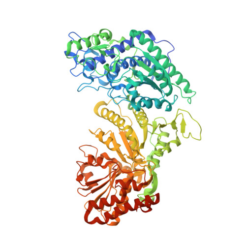
Crystal Structures of phosphoketolase: thiamine diphosphate-dependent dehydration mechanism

Suzuki, R.Katayama, T.Kim, B.-J.Wakagi, T.Shoun, H.Ashida, H.Yamamoto, K.Fushinobu, S.

(2010) J Biological Chem 285: 34279-34287

  • DOI: https://doi.org/10.1074/jbc.M110.156281
  • Primary Citation Related Structures: 
    3AHC, 3AHD, 3AHE, 3AHF, 3AHG, 3AHH, 3AHI, 3AHJ

  • PubMed Abstract: 

    Thiamine diphosphate (ThDP)-dependent enzymes are ubiquitously present in all organisms and catalyze essential reactions in various metabolic pathways. ThDP-dependent phosphoketolase plays key roles in the central metabolism of heterofermentative bacteria and in the pentose catabolism of various microbes. In particular, bifidobacteria, representatives of beneficial commensal bacteria, have an effective glycolytic pathway called bifid shunt in which 2.5 mol of ATP are produced per glucose. Phosphoketolase catalyzes two steps in the bifid shunt because of its dual-substrate specificity; they are phosphorolytic cleavage of fructose 6-phosphate or xylulose 5-phosphate to produce aldose phosphate, acetyl phosphate, and H(2)O. The phosphoketolase reaction is different from other well studied ThDP-dependent enzymes because it involves a dehydration step. Although phosphoketolase was discovered more than 50 years ago, its three-dimensional structure remains unclear. In this study we report the crystal structures of xylulose 5-phosphate/fructose 6-phosphate phosphoketolase from Bifidobacterium breve. The structures of the two intermediates before and after dehydration (α,β-dihydroxyethyl ThDP and 2-acetyl-ThDP) and complex with inorganic phosphate give an insight into the mechanism of each step of the enzymatic reaction.


  • Organizational Affiliation
    • Department of Biotechnology, The University of Tokyo, 1-1-1 Yayoi, Bunkyo-ku, Tokyo 113-8657, Japan.

Macromolecule Content 

  • Total Structure Weight: 95.73 kDa 
  • Atom Count: 7,192 
  • Modeled Residue Count: 802 
  • Deposited Residue Count: 845 
  • Unique protein chains: 1

Macromolecules

Find similar proteins by:|  3D Structure
Entity ID: 1
MoleculeChains  Sequence LengthOrganismDetailsImage
Xylulose 5-phosphate/fructose 6-phosphate phosphoketolase845Bifidobacterium breveMutation(s): 0 
Gene Names: xfp
EC: 4.1.2.22 (PDB Primary Data), 4.1.2 (UniProt)
UniProt
Find proteins for D6PAH1 (Bifidobacterium breve)
Explore D6PAH1 
Go to UniProtKB:  D6PAH1
Entity Groups
Sequence Clusters30% Identity50% Identity70% Identity90% Identity95% Identity100% Identity
UniProt GroupD6PAH1
Sequence Annotations
Expand
Reference Sequence

Small Molecules

Ligands 4 Unique
IDChains Name / Formula / InChI Key2D Diagram3D Interactions
HTL

Query on HTL


Download:Ideal Coordinates CCD File
C [auth A]2-ACETYL-THIAMINE DIPHOSPHATE
C14 H21 N4 O8 P2 S
GYRGKLZCJRVYRV-UHFFFAOYSA-O
EDO

Query on EDO


Download:Ideal Coordinates CCD File
E [auth A],
F [auth A],
G [auth A],
H [auth A]
1,2-ETHANEDIOL
C2 H6 O2
LYCAIKOWRPUZTN-UHFFFAOYSA-N
MG

Query on MG


Download:Ideal Coordinates CCD File
B [auth A]MAGNESIUM ION
Mg
JLVVSXFLKOJNIY-UHFFFAOYSA-N
NA

Query on NA


Download:Ideal Coordinates CCD File
D [auth A]SODIUM ION
Na
FKNQFGJONOIPTF-UHFFFAOYSA-N

Experimental Data & Validation

Experimental Data

  • Method: X-RAY DIFFRACTION
  • Resolution: 1.90 Å
  • R-Value Free:  0.197 (Depositor), 0.198 (DCC) 
  • R-Value Work:  0.162 (Depositor), 0.163 (DCC) 
  • R-Value Observed: 0.164 (Depositor) 
Space Group: I 4 2 2
Unit Cell:
Length ( Å )Angle ( ˚ )
a = 174.448α = 90
b = 174.448β = 90
c = 163.847γ = 90
Software Package:
Software NamePurpose
HKL-2000data collection
REFMACrefinement
HKL-2000data reduction
HKL-2000data scaling
REFMACphasing

Structure Validation

View Full Validation Report


Ligand Structure Quality Assessment 


Entry History 

Deposition Data

Revision History  (Full details and data files)

  • Version 1.0: 2010-08-25
    Type: Initial release
  • Version 1.1: 2011-07-13
    Changes: Version format compliance
  • Version 1.2: 2023-11-01
    Changes: Data collection, Database references, Derived calculations, Refinement description, Structure summary