


Binding mode of azide ion to pharaonis halorhodopsin
Kanada, S., Kouyama, T.To be published.
Experimental Data Snapshot
Starting Model: experimental
View more details
Entity ID: 1 | |||||
|---|---|---|---|---|---|
| Molecule | Chains | Sequence Length | Organism | Details | Image |
| Halorhodopsin | A, B, C [auth D] | 291 | Natronomonas pharaonis DSM 2160 | Mutation(s): 0 | 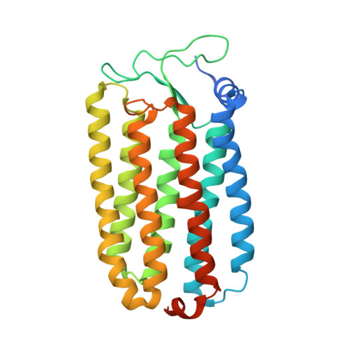 |
| Ligands 4 Unique | |||||
|---|---|---|---|---|---|
| ID | Chains | Name / Formula / InChI Key | 2D Diagram | 3D Interactions | |
| L3P Download:Ideal Coordinates CCD File | F [auth A] G [auth A] H [auth A] K [auth B] L [auth B] | 2,3-DI-O-PHYTANLY-3-SN-GLYCERO-1-PHOSPHORYL-3'-SN-GLYCEROL-1'-PHOSPHATE C46 H94 O11 P2 TZXJQSKPTCRGCA-VZSPAKCESA-L |  | ||
| 22B Download:Ideal Coordinates CCD File | E [auth A], N [auth B] | BACTERIORUBERIN C50 H76 O4 UVCQMCCIAHQDAF-CUMPQFAQSA-N |  | ||
| RET Download:Ideal Coordinates CCD File | D [auth A], J [auth B], P [auth D] | RETINAL C20 H28 O NCYCYZXNIZJOKI-OVSJKPMPSA-N |  | ||
| AZI Download:Ideal Coordinates CCD File | I [auth A], O [auth B], T [auth D] | AZIDE ION N3 IVRMZWNICZWHMI-UHFFFAOYSA-N |  | ||
| Length ( Å ) | Angle ( ˚ ) |
|---|---|
| a = 152.09 | α = 90 |
| b = 98.79 | β = 127.85 |
| c = 100.14 | γ = 90 |
| Software Name | Purpose |
|---|---|
| MAR345dtb | data collection |
| CNS | refinement |
| MOSFLM | data reduction |
| SCALA | data scaling |
| CNS | phasing |


