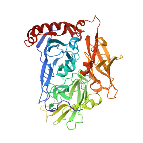
3ABG

X-ray Crystal Analysis of Bilirubin Oxidase from Myrothecium verrucaria at 2.3 angstrom Resolution using a Twin Crystal


Experimental Data Snapshot

  • Method: X-RAY DIFFRACTION
  • Resolution: 2.30 Å
  • R-Value Free: 0.186 
  • R-Value Work: 0.158 
  • R-Value Observed: 0.173 

wwPDB Validation   3D Report Full Report


This is version 3.0 of the entry. See complete history


Literature

X-ray analysis of bilirubin oxidase from Myrothecium verrucaria at 2.3 A resolution using a twinned crystal

Mizutani, K.Toyoda, M.Sagara, K.Takahashi, N.Sato, A.Kamitaka, Y.Tsujimura, S.Nakanishi, Y.Sugiura, T.Yamaguchi, S.Kano, K.Mikami, B.

(2010) Acta Crystallogr Sect F Struct Biol Cryst Commun 66: 765-770

  • DOI: https://doi.org/10.1107/S1744309110018828
  • Primary Citation of Related Structures:  
    3ABG

  • PubMed Abstract: 

    Bilirubin oxidase (BOD), a multicopper oxidase found in Myrothecium verrucaria, catalyzes the oxidation of bilirubin to biliverdin. Oxygen is the electron acceptor and is reduced to water. BOD is used for diagnostic analysis of bilirubin in serum and has attracted considerable attention as an enzymatic catalyst for the cathode of biofuel cells that work under neutral conditions. Here, the crystal structure of BOD is reported for the first time. Blue bipyramid-shaped crystals of BOD obtained in 2-methyl-2,4-pentanediol (MPD) and ammonium sulfate solution were merohedrally twinned in space group P6(3). Structure determination was achieved by the single anomalous diffraction (SAD) method using the anomalous diffraction of Cu atoms and synchrotron radiation and twin refinement was performed in the resolution range 33-2.3 A. The overall organization of BOD is almost the same as that of other multicopper oxidases: the protein is folded into three domains and a total of four copper-binding sites are found in domains 1 and 3. Although the four copper-binding sites were almost identical to those of other multicopper oxidases, the hydrophilic Asn residue (at the same position as a hydrophobic residue such as Leu in other multicopper oxidases) very close to the type I copper might contribute to the characteristically high redox potential of BOD.


  • Organizational Affiliation

    Laboratory of Applied Structural Biology, Division of Applied Life Sciences, The Graduate School of Agriculture, Kyoto University, Uji, Kyoto 611-0011, Japan. kmizutani@kais.kyoto-u.ac.jp


Macromolecules
Find similar proteins by:  (by identity cutoff)  |  3D Structure
Entity ID: 1
MoleculeChains Sequence LengthOrganismDetailsImage
Bilirubin oxidase
A, B
534Albifimbria verrucariaMutation(s): 0 
EC: 1.3.3.5
UniProt
Find proteins for Q12737 (Albifimbria verrucaria)
Explore Q12737 
Go to UniProtKB:  Q12737
Entity Groups  
Sequence Clusters30% Identity50% Identity70% Identity90% Identity95% Identity100% Identity
UniProt GroupQ12737
Sequence Annotations
Expand
  • Reference Sequence
Oligosaccharides

Help

Entity ID: 2
MoleculeChains Length2D Diagram Glycosylation3D Interactions
beta-D-mannopyranose-(1-4)-2-acetamido-2-deoxy-beta-D-glucopyranose-(1-4)-2-acetamido-2-deoxy-beta-D-glucopyranose
C
3N-Glycosylation
Glycosylation Resources
GlyTouCan:  G15407YE
GlyCosmos:  G15407YE
GlyGen:  G15407YE
Entity ID: 3
MoleculeChains Length2D Diagram Glycosylation3D Interactions
2-acetamido-2-deoxy-beta-D-glucopyranose-(1-4)-2-acetamido-2-deoxy-beta-D-glucopyranose
D, F
2N-Glycosylation
Glycosylation Resources
GlyTouCan:  G42666HT
GlyCosmos:  G42666HT
GlyGen:  G42666HT
Entity ID: 4
MoleculeChains Length2D Diagram Glycosylation3D Interactions
alpha-D-mannopyranose-(1-3)-alpha-D-mannopyranose-(1-6)-beta-D-mannopyranose-(1-4)-2-acetamido-2-deoxy-beta-D-glucopyranose-(1-4)-2-acetamido-2-deoxy-beta-D-glucopyranose
E
5N-Glycosylation
Glycosylation Resources
GlyTouCan:  G10756ZZ
GlyCosmos:  G10756ZZ
GlyGen:  G10756ZZ
Small Molecules
Ligands 3 Unique
IDChains Name / Formula / InChI Key2D Diagram3D Interactions
GOL
Query on GOL

Download Ideal Coordinates CCD File 
L [auth A]
M [auth A]
N [auth A]
O [auth A]
U [auth B]
L [auth A],
M [auth A],
N [auth A],
O [auth A],
U [auth B],
V [auth B],
W [auth B],
X [auth B]
GLYCEROL
C3 H8 O3
PEDCQBHIVMGVHV-UHFFFAOYSA-N
CU (Subject of Investigation/LOI)
Query on CU

Download Ideal Coordinates CCD File 
G [auth A]
H [auth A]
I [auth A]
J [auth A]
P [auth B]
G [auth A],
H [auth A],
I [auth A],
J [auth A],
P [auth B],
Q [auth B],
R [auth B],
S [auth B]
COPPER (II) ION
Cu
JPVYNHNXODAKFH-UHFFFAOYSA-N
OXY (Subject of Investigation/LOI)
Query on OXY

Download Ideal Coordinates CCD File 
K [auth A],
T [auth B]
OXYGEN MOLECULE
O2
MYMOFIZGZYHOMD-UHFFFAOYSA-N
Experimental Data & Validation

Experimental Data

  • Method: X-RAY DIFFRACTION
  • Resolution: 2.30 Å
  • R-Value Free: 0.186 
  • R-Value Work: 0.158 
  • R-Value Observed: 0.173 
  • Space Group: P 63
Unit Cell:
Length ( Å )Angle ( ˚ )
a = 139.742α = 90
b = 139.742β = 90
c = 135.561γ = 120
Software Package:
Software NamePurpose
PHENIXrefinement
HKL-2000data collection
SOLVEphasing
HKL-2000data reduction
HKL-2000data scaling

Structure Validation

View Full Validation Report


Entry History & Funding Information

Deposition Data


Funding OrganizationLocationGrant Number
Not funded--

Revision History  (Full details and data files)

  • Version 1.0: 2010-08-18
    Type: Initial release
  • Version 1.1: 2011-07-13
    Changes: Version format compliance
  • Version 2.0: 2020-07-29
    Type: Remediation
    Reason: Carbohydrate remediation
    Changes: Advisory, Atomic model, Data collection, Derived calculations, Non-polymer description, Structure summary
  • Version 3.0: 2023-07-26
    Changes: Advisory, Atomic model, Author supporting evidence, Data collection, Database references, Derived calculations, Non-polymer description, Other, Refinement description, Source and taxonomy, Structure summary