Activity and structure of the active-site mutants R386Y and R386F of Escherichia coli aspartate aminotransferase.
Danishefsky, A.T., Onnufer, J.J., Petsko, G.A., Ringe, D.(1991) Biochemistry 30: 1980-1985
- PubMed: 1993208 Search on PubMed
- DOI: https://doi.org/10.1021/bi00221a035
- Primary Citation Related Structures:
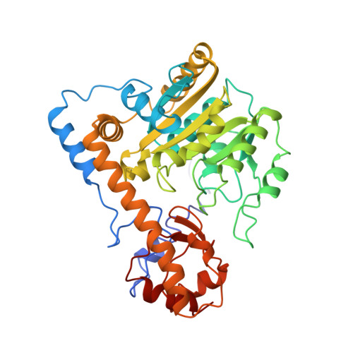
3AAT - PubMed Abstract:
Arginine-386, the active-site residue of Escherichia coli aspartate aminotransferase (EC 2.6.1.1) that binds the substrate alpha-carboxylate, was replaced with tyrosine and phenylalanine by site-directed mutagenesis. This experiment was undertaken to elucidate the roles of particular enzyme-substrate interactions in triggering the substrate-induced conformational change in the enzyme. The activity and crystal structure of the resulting mutants were examined. The apparent second-order rate constants of both of these mutants are reduced by more than 5 orders of magnitude as compared to that of wild-type enzyme, though R386Y is slightly more active than R386F. The 2.5-A resolution structure of R386F in its native state was determined by using difference Fourier methods. The overall structure is very similar to that of the wild-type enzyme in the open conformation. The position of the Phe-386 side chain, however, appears to shift with respect to that of Arg-386 in the wild-type enzyme and to form new contacts with neighboring residues.
- Department of Chemistry, Massachusetts Institute of Technology, Cambridge 02139.
Organizational Affiliation: