Remarkable improvement in the heat stability of CutA1 from Escherichia coli by rational protein design
Matsuura, Y., Ota, M., Tanaka, T., Takehira, M., Ogasahara, K., Bagautdinov, B., Kunishima, N., Yutani, K.(2010) J Biochem 148: 449-458
- PubMed: 20639520 Search on PubMed
- DOI: https://doi.org/10.1093/jb/mvq079
- Primary Citation Related Structures:
3AA8, 3AA9, 3AH6 - PubMed Abstract:
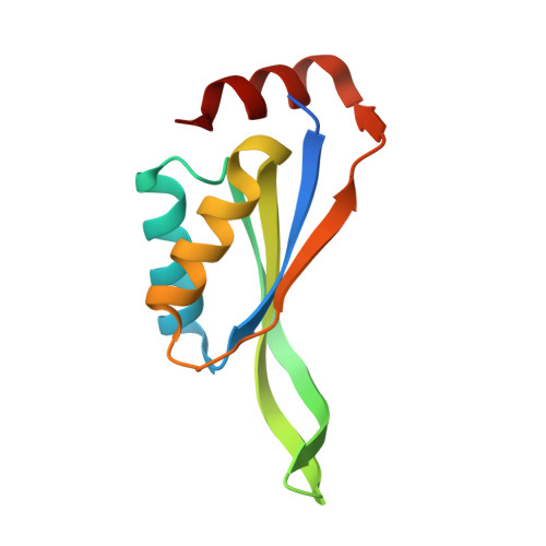
To enhance the heat stability of the CutA1 protein from Escherichia coli (EcCutA1) so that it has comparable stability to CutA1 from Pyrococcus horikoshii with a denaturation temperature (T(d)) of 150°C, we used the Stability Profile of Mutant Protein (SPMP) to examine the structure-sequence (3D-1D) compatibility between the conformation of EcCutA1 and its native sequence [J. Mol. Biol., 248, 733-738, (1995)]. We identified seven residues in EcCutA1 that were incompatible in terms of dihedral angles and hydrophobicity. These residues were replaced with appropriate amino acids, and the mutant proteins were evaluated for changes in stability by DSC and denaturant denaturation. The mutations that were introduced at five out of the seven positions improved the stability of EcCutA1. The T(d) values of single (S11A) and triple (S11V/E61V/Q73V) mutants improved by 16.5 and 26.6°C, respectively, compared to that of the wild-type protein (89.9°C). These analyses showed that (1) the stability of EcCutA1 is remarkably improved by slight substitutions, even though the stability of the wild-type protein is considerably high, (2) remarkable improvements in the stability can be quantitatively explained based on the newly solved native structure, and (3) SPMP is a powerful tool to examine substitutions that improve protein stability.
- RIKEN SPring-8 Center, Harima Institute, RIKEN; 1-1-1 Kouto, Sayo, Hyogo, Japan.
Organizational Affiliation: