3A5G

Crystal structure of a hemoglobin component V from Propsilocerus akamusi (pH7.0 coordinates)


Experimental Data Snapshot

  • Method: X-RAY DIFFRACTION
  • Resolution: 1.81 Å
  • R-Value Free: 0.208 
  • R-Value Work: 0.190 
  • R-Value Observed: 0.191 

wwPDB Validation   3D Report Full Report


Ligand Structure Quality Assessment 


This is version 1.2 of the entry. See complete history


Literature

pH-dependent structural changes in haemoglobin component V from the midge larva Propsilocerus akamusi (Orthocladiinae, Diptera)

Kuwada, T.Hasegawa, T.Takagi, T.Sato, I.Shishikura, F.

(2010) Acta Crystallogr D Biol Crystallogr 66: 258-267

  • DOI: https://doi.org/10.1107/S0907444909055760
  • Primary Citation of Related Structures:  
    2ZWJ, 3A5A, 3A5B, 3A5G, 3A9M

  • PubMed Abstract: 

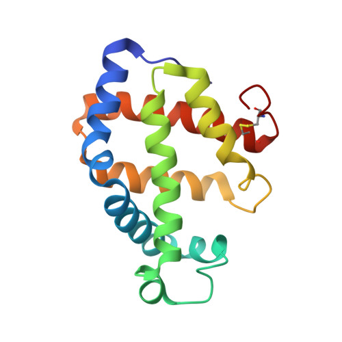
    Haemoglobin component V (Hb V) from the midge larva Propsilocerus akamusi exhibits oxygen affinity despite the replacement of HisE7 and a pH-dependence of its functional properties. In order to understand the contribution of the distal residue to the ligand-binding properties and the pH-dependent structural changes in this insect Hb, the crystal structure of Hb V was determined under five different pH conditions. Structural comparisons of these Hb structures indicated that at neutral pH ArgE10 contributes to the stabilization of the haem-bound ligand molecule as a functional substitute for the nonpolar E7 residue. However, ArgE10 does not contribute to stabilization at acidic and alkaline pH because of the swinging movement of the Arg side chain under these conditions. This pH-dependent behaviour of Arg results in significant differences in the hydrogen-bond network on the distal side of the haem in the Hb V structures at different pH values. Furthermore, the change in pH results in a partial movement of the F helix, considering that coupled movements of ArgE10 and the F helix determine the haem location at each pH. These results suggested that Hb V retains its functional properties by adapting to the structural changes caused by amino-acid replacements.


  • Organizational Affiliation

    Laboratory for Electron Beam Research and Application (LEBRA), Institute of Quantum Science, Nihon University, Funabashi, Chiba 274-8501, Japan. kuwadat@lebra.nihon-u.ac.jp


Macromolecules
Find similar proteins by:  (by identity cutoff)  |  3D Structure
Entity ID: 1
MoleculeChains Sequence LengthOrganismDetailsImage
Hemoglobin V152Tokunagayusurika akamusiMutation(s): 0 
UniProt
Find proteins for Q7M422 (Tokunagayusurika akamusi)
Explore Q7M422 
Go to UniProtKB:  Q7M422
Entity Groups  
Sequence Clusters30% Identity50% Identity70% Identity90% Identity95% Identity100% Identity
UniProt GroupQ7M422
Sequence Annotations
Expand
  • Reference Sequence
Small Molecules
Ligands 2 Unique
IDChains Name / Formula / InChI Key2D Diagram3D Interactions
HEM
Query on HEM

Download Ideal Coordinates CCD File 
B [auth A]PROTOPORPHYRIN IX CONTAINING FE
C34 H32 Fe N4 O4
KABFMIBPWCXCRK-RGGAHWMASA-L
CMO
Query on CMO

Download Ideal Coordinates CCD File 
C [auth A]CARBON MONOXIDE
C O
UGFAIRIUMAVXCW-UHFFFAOYSA-N
Experimental Data & Validation

Experimental Data

  • Method: X-RAY DIFFRACTION
  • Resolution: 1.81 Å
  • R-Value Free: 0.208 
  • R-Value Work: 0.190 
  • R-Value Observed: 0.191 
  • Space Group: P 21 21 21
Unit Cell:
Length ( Å )Angle ( ˚ )
a = 33.51α = 90
b = 64.14β = 90
c = 72.66γ = 90
Software Package:
Software NamePurpose
CrystalCleardata collection
CNXrefinement
CrystalCleardata reduction
CrystalCleardata scaling
CNXphasing

Structure Validation

View Full Validation Report


Ligand Structure Quality Assessment 


Entry History 

Deposition Data

Revision History  (Full details and data files)

  • Version 1.0: 2010-02-09
    Type: Initial release
  • Version 1.1: 2011-07-13
    Changes: Version format compliance
  • Version 1.2: 2023-11-01
    Changes: Data collection, Database references, Derived calculations, Refinement description