3A33

UbcH5b~Ubiquitin Conjugate


Experimental Data Snapshot

  • Method: X-RAY DIFFRACTION
  • Resolution: 2.20 Å
  • R-Value Free: 0.280 
  • R-Value Work: 0.228 
  • R-Value Observed: 0.231 

wwPDB Validation   3D Report Full Report


This is version 1.2 of the entry. See complete history


Literature

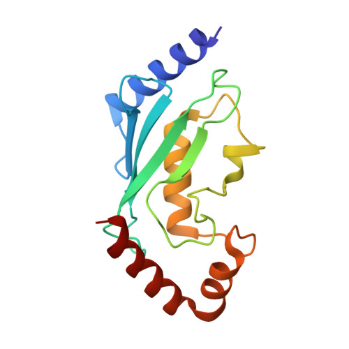
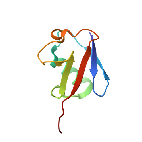
Crystal Structure of UbcH5b~Ubiquitin Intermediate: Insight into the Formation of the Self-Assembled E2~Ub Conjugates

Sakata, E.Satoh, T.Yamamoto, S.Yamaguchi, Y.Yagi-Utsumi, M.Kurimoto, E.Tanaka, K.Wakatsuki, S.Kato, K.

(2010) Structure 18: 138-147

  • DOI: https://doi.org/10.1016/j.str.2009.11.007
  • Primary Citation of Related Structures:  
    3A33

  • PubMed Abstract: 

    E2 ubiquitin-conjugating enzymes catalyze the attachment of ubiquitin to lysine residues of target proteins. The UbcH5b E2 enzyme has been shown to play a key role in the initiation of the ubiquitination of substrate proteins upon action of several E3 ligases. Here we have determined the 2.2 A crystal structure of an intermediate of UbcH5b~ubiquitin (Ub) conjugate, which is assembled into an infinite spiral through the backside interaction. This active complex may provide multiple E2 active sites, enabling efficient ubiquitination of substrates. Indeed, biochemical assays support a model in which the self-assembled UbcH5b~Ub can serve as a bridge for the gap between the lysine residue of the substrate and the catalytic cysteine of E2.


  • Organizational Affiliation

    Department of Structural Biology and Biomolecular Engineering, Graduate School of Pharmaceutical Sciences, Nagoya City University, 3-1 Tanabe-dori, Mizuho-ku, Nagoya 467-8603, Japan.


Macromolecules
Find similar proteins by:  (by identity cutoff)  |  3D Structure
Entity ID: 1
MoleculeChains Sequence LengthOrganismDetailsImage
Ubiquitin-conjugating enzyme E2 D2150Homo sapiensMutation(s): 0 
Gene Names: UBCH5B
EC: 6.3.2.19
UniProt & NIH Common Fund Data Resources
Find proteins for P62837 (Homo sapiens)
Explore P62837 
Go to UniProtKB:  P62837
PHAROS:  P62837
GTEx:  ENSG00000131508 
Entity Groups  
Sequence Clusters30% Identity50% Identity70% Identity90% Identity95% Identity100% Identity
UniProt GroupP62837
Sequence Annotations
Expand
  • Reference Sequence
Find similar proteins by:  (by identity cutoff)  |  3D Structure
Entity ID: 2
MoleculeChains Sequence LengthOrganismDetailsImage
Ubiquitin76Homo sapiensMutation(s): 0 
UniProt & NIH Common Fund Data Resources
Find proteins for P0CG48 (Homo sapiens)
Explore P0CG48 
Go to UniProtKB:  P0CG48
PHAROS:  P0CG48
GTEx:  ENSG00000150991 
Entity Groups  
Sequence Clusters30% Identity50% Identity70% Identity90% Identity95% Identity100% Identity
UniProt GroupP0CG48
Sequence Annotations
Expand
  • Reference Sequence
Small Molecules
Ligands 1 Unique
IDChains Name / Formula / InChI Key2D Diagram3D Interactions
GOL
Query on GOL

Download Ideal Coordinates CCD File 
C [auth A]GLYCEROL
C3 H8 O3
PEDCQBHIVMGVHV-UHFFFAOYSA-N
Experimental Data & Validation

Experimental Data

  • Method: X-RAY DIFFRACTION
  • Resolution: 2.20 Å
  • R-Value Free: 0.280 
  • R-Value Work: 0.228 
  • R-Value Observed: 0.231 
  • Space Group: P 61 2 2
Unit Cell:
Length ( Å )Angle ( ˚ )
a = 72.5α = 90
b = 72.5β = 90
c = 176.6γ = 120
Software Package:
Software NamePurpose
REFMACrefinement
HKL-2000data collection
DENZOdata reduction
SCALEPACKdata scaling
MOLREPphasing

Structure Validation

View Full Validation Report


Entry History 

Deposition Data

Revision History  (Full details and data files)

  • Version 1.0: 2009-11-24
    Type: Initial release
  • Version 1.1: 2011-07-13
    Changes: Non-polymer description, Version format compliance
  • Version 1.2: 2023-11-01
    Changes: Data collection, Database references, Derived calculations, Refinement description