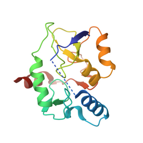
3A1B

Crystal structure of the DNMT3A ADD domain in complex with histone H3


Experimental Data Snapshot

  • Method: X-RAY DIFFRACTION
  • Resolution: 2.29 Å
  • R-Value Free: 0.224 
  • R-Value Work: 0.192 
  • R-Value Observed: 0.193 

Starting Model: experimental
View more details

wwPDB Validation   3D Report Full Report


This is version 1.3 of the entry. See complete history


Literature

Structural basis for recognition of H3K4 methylation status by the DNA methyltransferase 3A ATRX-DNMT3-DNMT3L domain

Otani, J.Nankumo, T.Arita, K.Inamoto, S.Ariyoshi, M.Shirakawa, M.

(2009) EMBO Rep 10: 1235-1241

  • DOI: https://doi.org/10.1038/embor.2009.218
  • Primary Citation of Related Structures:  
    3A1A, 3A1B

  • PubMed Abstract: 

    DNMT3 proteins are de novo DNA methyltransferases that are responsible for the establishment of DNA methylation patterns in mammalian genomes. Here, we have determined the crystal structures of the ATRX-DNMT3-DNMT3L (ADD) domain of DNMT3A in an unliganded form and in a complex with the amino-terminal tail of histone H3. Combined with the results of biochemical analysis, the complex structure indicates that DNMT3A recognizes the unmethylated state of lysine 4 in histone H3. This finding indicates that the recruitment of DNMT3A onto chromatin, and thereby de novo DNA methylation, is mediated by recognition of the histone modification state by its ADD domain. Furthermore, our biochemical and nuclear magnetic resonance data show mutually exclusive binding of the ADD domain of DNMT3A and the chromodomain of heterochromatin protein 1alpha to the H3 tail. These results indicate that de novo DNA methylation by DNMT3A requires the alteration of chromatin structure.


  • Organizational Affiliation

    Department of Molecular Engineering, Graduate School of Engineering, Kyoto University, Kyoto-Daigaku Katsura, Nishikyo-Ku, Kyoto 615-8510, Japan.


Macromolecules
Find similar proteins by:  (by identity cutoff)  |  3D Structure
Entity ID: 1
MoleculeChains Sequence LengthOrganismDetailsImage
DNA (cytosine-5)-methyltransferase 3A, Histone H3.1159Homo sapiensMutation(s): 0 
EC: 2.1.1.37
UniProt & NIH Common Fund Data Resources
Find proteins for P68431 (Homo sapiens)
Explore P68431 
Go to UniProtKB:  P68431
PHAROS:  P68431
Find proteins for Q9Y6K1 (Homo sapiens)
Explore Q9Y6K1 
Go to UniProtKB:  Q9Y6K1
PHAROS:  Q9Y6K1
GTEx:  ENSG00000119772 
Entity Groups  
Sequence Clusters30% Identity50% Identity70% Identity90% Identity95% Identity100% Identity
UniProt GroupsP68431Q9Y6K1
Sequence Annotations
Expand
  • Reference Sequence
Small Molecules
Ligands 2 Unique
IDChains Name / Formula / InChI Key2D Diagram3D Interactions
ZN
Query on ZN

Download Ideal Coordinates CCD File 
B [auth A],
C [auth A],
D [auth A]
ZINC ION
Zn
PTFCDOFLOPIGGS-UHFFFAOYSA-N
EDO
Query on EDO

Download Ideal Coordinates CCD File 
E [auth A]
F [auth A]
G [auth A]
H [auth A]
I [auth A]
E [auth A],
F [auth A],
G [auth A],
H [auth A],
I [auth A],
J [auth A],
K [auth A]
1,2-ETHANEDIOL
C2 H6 O2
LYCAIKOWRPUZTN-UHFFFAOYSA-N
Experimental Data & Validation

Experimental Data

  • Method: X-RAY DIFFRACTION
  • Resolution: 2.29 Å
  • R-Value Free: 0.224 
  • R-Value Work: 0.192 
  • R-Value Observed: 0.193 
  • Space Group: P 41 21 2
Unit Cell:
Length ( Å )Angle ( ˚ )
a = 68.547α = 90
b = 68.547β = 90
c = 109.95γ = 90
Software Package:
Software NamePurpose
REFMACrefinement
ADSCdata collection
HKL-2000data reduction
HKL-2000data scaling
MOLREPphasing

Structure Validation

View Full Validation Report


Entry History 

Deposition Data

Revision History  (Full details and data files)

  • Version 1.0: 2009-11-10
    Type: Initial release
  • Version 1.1: 2011-07-13
    Changes: Advisory, Version format compliance
  • Version 1.2: 2017-08-09
    Changes: Advisory, Source and taxonomy
  • Version 1.3: 2023-11-01
    Changes: Advisory, Data collection, Database references, Derived calculations, Refinement description