3A18

Crystal Structure of Aldoxime Dehydratase (OxdRE) in Complex with Butyraldoxime (soaked crystal)


Experimental Data Snapshot

  • Method: X-RAY DIFFRACTION
  • Resolution: 1.80 Å
  • R-Value Free: 0.280 
  • R-Value Work: 0.247 
  • R-Value Observed: 0.249 

wwPDB Validation   3D Report Full Report


This is version 1.3 of the entry. See complete history


Literature

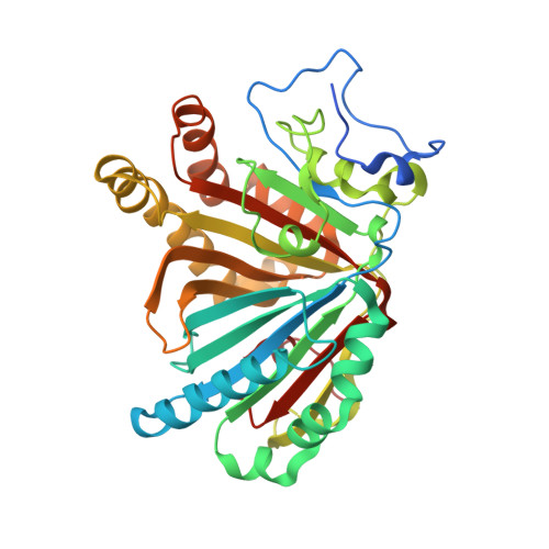
X-ray crystal structure of michaelis complex of aldoxime dehydratase

Sawai, H.Sugimoto, H.Kato, Y.Asano, Y.Shiro, Y.Aono, S.

(2009) J Biol Chem 284: 32089-32096

  • DOI: https://doi.org/10.1074/jbc.M109.018762
  • Primary Citation of Related Structures:  
    3A15, 3A16, 3A17, 3A18

  • PubMed Abstract: 

    Aldoxime dehydratase (Oxd) catalyzes the dehydration of aldoximes (R-CH=N-OH) to their corresponding nitrile (R-C triple bond N). Oxd is a heme-containing enzyme that catalyzes the dehydration reaction as its physiological function. We have determined the first two structures of Oxd: the substrate-free OxdRE at 1.8 A resolution and the n-butyraldoxime- and propionaldoxime-bound OxdREs at 1.8 and 1.6 A resolutions, respectively. Unlike other heme enzymes, the organic substrate is directly bound to the heme iron in OxdRE. We determined the structure of the Michaelis complex of OxdRE by using the unique substrate binding and activity regulation properties of Oxd. The Michaelis complex was prepared by x-ray cryoradiolytic reduction of the ferric dead-end complex in which Oxd contains a Fe(3+) heme form. The crystal structures reveal the mechanism of substrate recognition and the catalysis of OxdRE.


  • Organizational Affiliation

    From the Okazaki Institute for Integrative Bioscience, National Institutes of Natural Sciences, 5-1 Higashiyama, Myodaiji, Okazaki 444-8787, Japan.


Macromolecules
Find similar proteins by:  (by identity cutoff)  |  3D Structure
Entity ID: 1
MoleculeChains Sequence LengthOrganismDetailsImage
Aldoxime dehydratase
A, B, C, D
373Rhodococcus erythropolisMutation(s): 0 
Gene Names: oxd
EC: 4.99.1.5
UniProt
Find proteins for Q76K71 (Rhodococcus erythropolis)
Explore Q76K71 
Go to UniProtKB:  Q76K71
Entity Groups  
Sequence Clusters30% Identity50% Identity70% Identity90% Identity95% Identity100% Identity
UniProt GroupQ76K71
Sequence Annotations
Expand
  • Reference Sequence
Experimental Data & Validation

Experimental Data

  • Method: X-RAY DIFFRACTION
  • Resolution: 1.80 Å
  • R-Value Free: 0.280 
  • R-Value Work: 0.247 
  • R-Value Observed: 0.249 
  • Space Group: P 1 21 1
Unit Cell:
Length ( Å )Angle ( ˚ )
a = 63.118α = 90
b = 148.569β = 90.3
c = 79.113γ = 90
Software Package:
Software NamePurpose
DENZOdata reduction
SCALEPACKdata scaling
MOLREPphasing
REFMACrefinement
PDB_EXTRACTdata extraction
SPACEdata collection
HKL-2000data reduction
HKL-2000data scaling

Structure Validation

View Full Validation Report


Entry History 

Deposition Data

Revision History  (Full details and data files)

  • Version 1.0: 2009-09-08
    Type: Initial release
  • Version 1.1: 2011-07-13
    Changes: Advisory, Refinement description, Version format compliance
  • Version 1.2: 2017-10-11
    Changes: Refinement description
  • Version 1.3: 2023-11-01
    Changes: Data collection, Database references, Derived calculations, Refinement description