3A0T | pdb_00003a0t

Catalytic domain of histidine kinase ThkA (TM1359) in complex with ADP and Mg ion (trigonal)


Experimental Data Snapshot

  • Method: X-RAY DIFFRACTION
  • Resolution: 1.91 Å
  • R-Value Free: 
    0.213 (Depositor), 0.209 (DCC) 
  • R-Value Work: 
    0.185 (Depositor), 0.182 (DCC) 
  • R-Value Observed: 
    0.186 (Depositor) 

Starting Model: other
View more details

wwPDB Validation 3D Report Full Report

Validation slider image for 3A0T

Ligand Structure Quality Assessment 


This is version 1.4 of the entry. See complete history

Literature

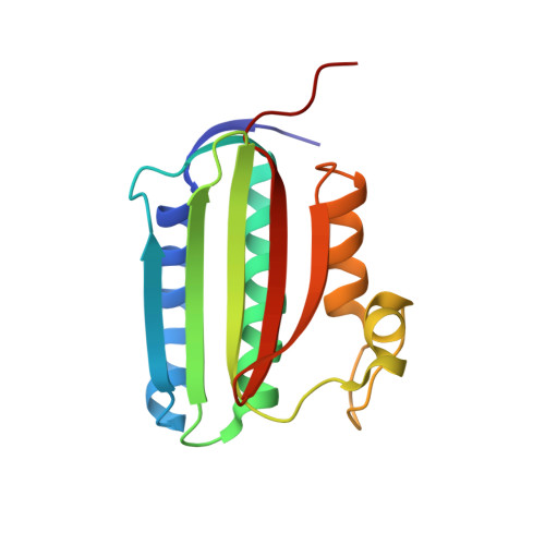
Structure of PAS-linked histidine kinase and the response regulator complex

Yamada, S.Sugimoto, H.Kobayashi, M.Ohno, A.Nakamura, H.Shiro, Y.

(2009) Structure 17: 1333-1344

  • DOI: https://doi.org/10.1016/j.str.2009.07.016
  • Primary Citation Related Structures: 
    3A0R, 3A0S, 3A0T, 3A0U, 3A0V, 3A0W, 3A0X, 3A0Y, 3A0Z, 3A10

  • PubMed Abstract: 

    We determined the structure of the complex of the sensory histidine kinase (HK) and its cognate response regulator (RR) in the two-component signal transduction system of Thermotoga maritima. This was accomplished by fitting the high-resolution structures of the isolated HK domains and the RR onto the electron density map (3.8 A resolution) of the HK/RR complex crystal. Based on the structural information, we evaluated the roles of both interdomain and intermolecular interactions in the signal transduction of the cytosolic PAS-linked HK and RR system, in particular the O(2)-sensor FixL/FixJ system. The PAS-sensor domain of HK interacts with the catalytic domain of the same polypeptide chain by creating an interdomain beta sheet. The interaction site between HK and RR, which was confirmed by NMR, is suitable for the intermolecular transfer reaction of the phosphoryl group, indicating that the observed interaction is important for the phosphatase activity of HK that dephosphorylates phospho-RR.


  • Organizational Affiliation
    • RIKEN SPring-8 Center, Kouto 1-1-1, Sayo, Hyogo, Japan.

Macromolecule Content 

  • Total Structure Weight: 18.18 kDa 
  • Atom Count: 1,444 
  • Modeled Residue Count: 152 
  • Deposited Residue Count: 152 
  • Unique protein chains: 1

Macromolecules

Find similar proteins by:|  3D Structure
Entity ID: 1
MoleculeChains  Sequence LengthOrganismDetailsImage
Sensor protein152Thermotoga maritimaMutation(s): 0 
Gene Names: TM_1359
EC: 2.7.13.3
UniProt
Find proteins for Q9X180 (Thermotoga maritima (strain ATCC 43589 / DSM 3109 / JCM 10099 / NBRC 100826 / MSB8))
Explore Q9X180 
Go to UniProtKB:  Q9X180
Entity Groups
Sequence Clusters30% Identity50% Identity70% Identity90% Identity95% Identity100% Identity
UniProt GroupQ9X180
Sequence Annotations
Expand
Reference Sequence

Small Molecules

Experimental Data & Validation

Experimental Data

  • Method: X-RAY DIFFRACTION
  • Resolution: 1.91 Å
  • R-Value Free:  0.213 (Depositor), 0.209 (DCC) 
  • R-Value Work:  0.185 (Depositor), 0.182 (DCC) 
  • R-Value Observed: 0.186 (Depositor) 
Space Group: P 32 2 1
Unit Cell:
Length ( Å )Angle ( ˚ )
a = 72.943α = 90
b = 72.943β = 90
c = 91.258γ = 120
Software Package:
Software NamePurpose
REFMACrefinement
PDB_EXTRACTdata extraction
HKL-2000data reduction
HKL-2000data scaling
REFMACphasing

Structure Validation

View Full Validation Report


Ligand Structure Quality Assessment 


Entry History 

Deposition Data

Revision History  (Full details and data files)

  • Version 1.0: 2009-10-20
    Type: Initial release
  • Version 1.1: 2011-07-13
    Changes: Version format compliance
  • Version 1.2: 2013-07-10
    Changes: Other
  • Version 1.3: 2024-03-13
    Changes: Data collection, Database references, Derived calculations
  • Version 1.4: 2024-04-03
    Changes: Refinement description