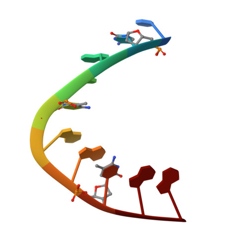
383D

Hydration and recognition of methylated CPG steps in DNA


Experimental Data Snapshot

  • Method: X-RAY DIFFRACTION
  • Resolution: 1.70 Å
  • R-Value Work: 0.196 
  • R-Value Observed: 0.196 

wwPDB Validation   3D Report Full Report


This is version 1.4 of the entry. See complete history


Literature

Hydration and Recognition of Methylated Cpg Steps in DNA

Mayer-Jung, C.Moras, D.Timsit, Y.

(1998) EMBO J 17: 2709

  • DOI: https://doi.org/10.1093/emboj/17.9.2709
  • Primary Citation of Related Structures:  
    382D, 383D, 384D

  • PubMed Abstract: 

    The analysis of the hydration pattern around methylated CpG steps in three high resolution (1.7, 2.15 and 2.2 A) crystal structures of A-DNA decamers reveals that the methyl groups of cytosine residues are well hydrated. In comparing the native structure with two structurally distinct forms of the decamer d(CCGCCGGCGG) fully methylated at its CpG steps, this study shows also that in certain structural and sequence contexts, the methylated cytosine base can be more hydrated that the unmodified one. These water molecules seem to be stabilized in front of the methyl group through the formation C-H...O interactions. In addition, these structures provide the first observation of magnesium cations bound to the major groove of A-DNA and reveal two distinct modes of metal binding in methylated and native duplexes. These findings suggest that methylated cytosine bases could be recognized by protein or DNA polar residues through their tightly bound water molecules.


  • Organizational Affiliation

    Laboratoire de Biologie Structurale IGBMC CNRS/INSERM/ULP, Parc d'Innovation, 1, rue Laurent Fries, Illkirch 67404, France.


Macromolecules

Find similar nucleic acids by:  Sequence   |   3D Structure  

Entity ID: 1
MoleculeChains LengthOrganismImage
DNA (5'-D(*CP*(5CM)P*GP*CP*(5CM)P*GP*GP*(5CM)P*GP*G)-3')
A, B
10N/A
Sequence Annotations
Expand
  • Reference Sequence
Small Molecules
Ligands 1 Unique
IDChains Name / Formula / InChI Key2D Diagram3D Interactions
MG
Query on MG

Download Ideal Coordinates CCD File 
C [auth B]MAGNESIUM ION
Mg
JLVVSXFLKOJNIY-UHFFFAOYSA-N
Experimental Data & Validation

Experimental Data

  • Method: X-RAY DIFFRACTION
  • Resolution: 1.70 Å
  • R-Value Work: 0.196 
  • R-Value Observed: 0.196 
  • Space Group: P 21 21 21
Unit Cell:
Length ( Å )Angle ( ˚ )
a = 23.9α = 90
b = 45.3β = 90
c = 46.9γ = 90
Software Package:
Software NamePurpose
XDSdata scaling
AUTOMARdata reduction
X-PLORmodel building
X-PLORrefinement
XDSdata reduction
X-PLORphasing

Structure Validation

View Full Validation Report


Entry History 

Deposition Data

Revision History  (Full details and data files)

  • Version 1.0: 1998-04-08
    Type: Initial release
  • Version 1.1: 2008-04-26
    Changes: Version format compliance
  • Version 1.2: 2011-07-13
    Changes: Version format compliance
  • Version 1.3: 2017-10-04
    Changes: Refinement description
  • Version 1.4: 2023-12-27
    Changes: Data collection, Database references, Derived calculations