348D

X-RAY CRYSTAL STRUCTURES OF THE DECAMER DGACCGCGGTC: LOW SALT CONCENTRATION

  • Classification: DNA
  • Mutation(s): No 

  • Deposited: 1997-08-29 Released: 1997-10-09 
  • Deposition Author(s): Luo, M., Finley, J.B.

Experimental Data Snapshot

  • Method: X-RAY DIFFRACTION
  • Resolution: 1.70 Å
  • R-Value Free: 0.210 
  • R-Value Work: 0.190 
  • R-Value Observed: 0.190 

wwPDB Validation   3D Report Full Report


This is version 1.3 of the entry. See complete history


Literature

X-ray crystal structures of half the human papilloma virus E2 binding site: d(GACCGCGGTC).

Finley, J.B.Luo, M.

(1998) Nucleic Acids Res 26: 5719-5727

  • DOI: https://doi.org/10.1093/nar/26.24.5719
  • Primary Citation of Related Structures:  
    348D, 349D, 401D

  • PubMed Abstract: 

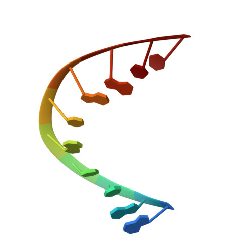
    The X-ray crystal structure of the DNA decamer d(GACCGCGGTC), containing half the human papilloma virus E2 binding site, has been solved from two crystals grown at different ionic conditions (50 mM MgCl2and 50 mM spermine or 1.56 mM MgCl2and 1.56 mM spermine). Despite the variation in salt concentration, the two DNA structures are in a very similar, A-type DNA conformation, with helical axes curving towards the major groove. Although the salt concentrations do not effect the helical parameters or hydration to a large degree, there is a change in the overall helical curvature; 18 degrees and 31 degrees for the low and high salt structures, respectively. This curvature appears to be sequence specific and biologically relevant when compared with similar DNA structures, including the E2 binding site of a protein-DNA complex.


  • Organizational Affiliation

    Center for Macromolecular Crystallography, Department of Microbiology, University of Alabama-Birmingham, Birmingham, AL 35294-0005, USA. jfinley@orion.cmc.uab.edu


Macromolecules

Find similar nucleic acids by:  Sequence   |   3D Structure  

Entity ID: 1
MoleculeChains LengthOrganismImage
DNA (5'-D(*GP*AP*CP*CP*GP*CP*GP*GP*TP*C)-3')10N/A
Sequence Annotations
Expand
  • Reference Sequence
Experimental Data & Validation

Experimental Data

  • Method: X-RAY DIFFRACTION
  • Resolution: 1.70 Å
  • R-Value Free: 0.210 
  • R-Value Work: 0.190 
  • R-Value Observed: 0.190 
  • Space Group: P 61 2 2
Unit Cell:
Length ( Å )Angle ( ˚ )
a = 38.606α = 90
b = 38.606β = 90
c = 80.573γ = 120
Software Package:
Software NamePurpose
X-PLORrefinement
XENGENdata reduction

Structure Validation

View Full Validation Report


Entry History 

Deposition Data

Revision History  (Full details and data files)

  • Version 1.0: 1997-10-09
    Type: Initial release
  • Version 1.1: 2008-05-22
    Changes: Version format compliance
  • Version 1.2: 2011-07-13
    Changes: Version format compliance
  • Version 1.3: 2024-02-21
    Changes: Data collection, Database references