333D | pdb_0000333d

THE CRYSTAL STRUCTURE OF AN RNA OLIGOMER INCORPORATING TANDEM ADENOSINE-INOSINE MISMATCHES


Experimental Data Snapshot

  • Method: X-RAY DIFFRACTION
  • Resolution: 2.52 Å
  • R-Value Free: 
    0.266 (Depositor) 
  • R-Value Work: 
    0.246 (Depositor), 0.254 (DCC) 
  • R-Value Observed: 
    0.246 (Depositor) 

Starting Model: experimental
View more details

wwPDB Validation 3D Report Full Report

Validation slider image for 333D

This is version 2.1 of the entry. See complete history

Literature

The crystal structure of an RNA oligomer incorporating tandem adenosine-inosine mismatches.

Carter, R.J.Baeyens, K.J.SantaLucia, J.Turner, D.H.Holbrook, S.R.

(1997) Nucleic Acids Res 25: 4117-4122

  • DOI: https://doi.org/10.1093/nar/25.20.4117
  • Primary Citation Related Structures: 
    333D

  • PubMed Abstract: 

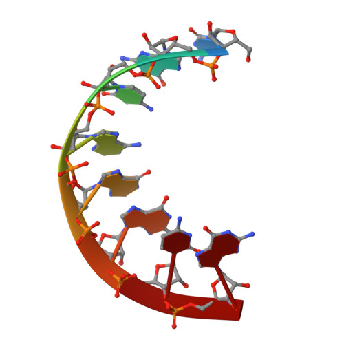
    The X-ray crystallographic structure of the RNA duplex [r(CGCAIGCG)]2 has been refined to 2.5 A. It shows a symmetric internal loop of two non-Watson-Crick base pairs which form in the middle of the duplex. The tandem A-I/I-A pairs are related by a crystallographic two-fold axis. Both A(anti)-I(anti) mismatches are in a head-to-head conformation forming hydrogen bonds using the Watson-Crick positions. The octamer duplexes stack above one another in the cell forming a pseudo-infinite helix throughout the crystal. A hydrated calcium ion bridges between the 3'-terminal of one molecule and the backbone of another. The tandem A-I mismatches are incorporated with only minor distortion to the backbone. This is in contrast to the large helical perturbations often produced by sheared G-A pairs in RNA oligonucleotides.


  • Organizational Affiliation
    • Structural Biology Division, Lawrence Berkeley Laboratory, Berkeley, CA 94720, USA.

Macromolecule Content 

  • Total Structure Weight: 2.61 kDa 
  • Atom Count: 177 
  • Modeled Residue Count: 8 
  • Deposited Residue Count: 8 
  • Unique nucleic acid chains: 1

Macromolecules

Find similar nucleic acids by:  Sequence
Entity ID: 1
MoleculeChains LengthOrganismImage
RNA (5'-R(*CP*GP*CP*AP*IP*GP*CP*G)-3')8N/A
Sequence Annotations
Expand
Reference Sequence

Small Molecules

Ligands 1 Unique
IDChains Name / Formula / InChI Key2D Diagram3D Interactions
CA

Query on CA


Download:Ideal Coordinates CCD File
B [auth A]CALCIUM ION
Ca
BHPQYMZQTOCNFJ-UHFFFAOYSA-N

Experimental Data & Validation

Experimental Data

  • Method: X-RAY DIFFRACTION
  • Resolution: 2.52 Å
  • R-Value Free:  0.266 (Depositor) 
  • R-Value Work:  0.246 (Depositor), 0.254 (DCC) 
  • R-Value Observed: 0.246 (Depositor) 
Space Group: P 61 2 2
Unit Cell:
Length ( Å )Angle ( ˚ )
a = 39.05α = 90
b = 39.05β = 90
c = 58.85γ = 120
Software Package:
Software NamePurpose
X-PLORmodel building
X-PLORrefinement
R-AXISdata reduction
X-PLORphasing

Structure Validation

View Full Validation Report


Entry History 

Deposition Data

Revision History  (Full details and data files)

  • Version 1.0: 1997-10-16
    Type: Initial release
  • Version 1.1: 2008-05-22
    Changes: Version format compliance
  • Version 1.2: 2011-07-13
    Changes: Version format compliance
  • Version 2.0: 2024-02-21
    Changes: Atomic model, Data collection, Database references, Derived calculations
  • Version 2.1: 2024-04-03
    Changes: Refinement description