2ZKM | pdb_00002zkm

Crystal Structure of Phospholipase C Beta 2


Experimental Data Snapshot

  • Method: X-RAY DIFFRACTION
  • Resolution: 1.62 Å
  • R-Value Free: 
    0.213 (Depositor), 0.212 (DCC) 
  • R-Value Work: 
    0.196 (Depositor), 0.195 (DCC) 
  • R-Value Observed: 
    0.197 (Depositor) 

Starting Model: experimental
View more details

wwPDB Validation 3D Report Full Report

Validation slider image for 2ZKM

This is version 1.3 of the entry. See complete history

Literature

General and versatile autoinhibition of PLC isozymes

Hicks, S.N.Jezyk, M.R.Gershburg, S.Seifert, J.P.Harden, T.K.Sondek, J.

(2008) Mol Cell 31: 383-394

  • DOI: https://doi.org/10.1016/j.molcel.2008.06.018
  • Primary Citation Related Structures: 
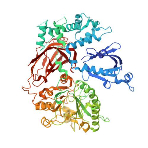
    2ZKM

  • PubMed Abstract: 

    Phospholipase C (PLC) isozymes are directly activated by heterotrimeric G proteins and Ras-like GTPases to hydrolyze phosphatidylinositol 4,5-bisphosphate into the second messengers diacylglycerol and inositol 1,4,5-trisphosphate. Although PLCs play central roles in myriad signaling cascades, the molecular details of their activation remain poorly understood. As described here, the crystal structure of PLC-beta2 illustrates occlusion of the active site by a loop separating the two halves of the catalytic TIM barrel. Removal of this insertion constitutively activates PLC-beta2 without ablating its capacity to be further stimulated by classical G protein modulators. Similar regulation occurs in other PLC members, and a general mechanism of interfacial activation at membranes is presented that provides a unifying framework for PLC activation by diverse stimuli.


  • Organizational Affiliation
    • Department of Pharmacology, The University of North Carolina School of Medicine, Chapel Hill, NC 27599, USA.

Macromolecule Content 

  • Total Structure Weight: 91.07 kDa 
  • Atom Count: 6,397 
  • Modeled Residue Count: 708 
  • Deposited Residue Count: 799 
  • Unique protein chains: 1

Macromolecules

Find similar proteins by:|  3D Structure
Entity ID: 1
MoleculeChains  Sequence LengthOrganismDetailsImage
1-phosphatidylinositol-4,5-bisphosphate phosphodiesterase beta-2A [auth X]799Homo sapiensMutation(s): 0 
Gene Names: PLC beta 2
EC: 3.1.4.11
UniProt & NIH Common Fund Data Resources
Find proteins for Q00722 (Homo sapiens)
Explore Q00722 
Go to UniProtKB:  Q00722
PHAROS:  Q00722
GTEx:  ENSG00000137841 
Entity Groups
Sequence Clusters30% Identity50% Identity70% Identity90% Identity95% Identity100% Identity
UniProt GroupQ00722
Sequence Annotations
Expand
Reference Sequence

Small Molecules

Ligands 1 Unique
IDChains Name / Formula / InChI Key2D Diagram3D Interactions
CA

Query on CA


Download:Ideal Coordinates CCD File
B [auth X]CALCIUM ION
Ca
BHPQYMZQTOCNFJ-UHFFFAOYSA-N

Experimental Data & Validation

Experimental Data

  • Method: X-RAY DIFFRACTION
  • Resolution: 1.62 Å
  • R-Value Free:  0.213 (Depositor), 0.212 (DCC) 
  • R-Value Work:  0.196 (Depositor), 0.195 (DCC) 
  • R-Value Observed: 0.197 (Depositor) 
Space Group: P 21 21 21
Unit Cell:
Length ( Å )Angle ( ˚ )
a = 80.404α = 90
b = 86.379β = 90
c = 147.447γ = 90
Software Package:
Software NamePurpose
DENZOdata reduction
SCALEPACKdata scaling
PHASERphasing
REFMACrefinement
PDB_EXTRACTdata extraction

Structure Validation

View Full Validation Report


Entry History 

Deposition Data

Revision History  (Full details and data files)

  • Version 1.0: 2008-08-12
    Type: Initial release
  • Version 1.1: 2011-07-13
    Changes: Version format compliance
  • Version 1.2: 2017-10-11
    Changes: Refinement description
  • Version 1.3: 2023-11-01
    Changes: Data collection, Database references, Derived calculations, Refinement description