2Z8L

Crystal Structure of the Staphylococcal superantigen-like protein SSL5 at pH 4.6 complexed with sialyl Lewis X


Experimental Data Snapshot

  • Method: X-RAY DIFFRACTION
  • Resolution: 1.65 Å
  • R-Value Free: 0.218 
  • R-Value Work: 0.181 
  • R-Value Observed: 0.183 

wwPDB Validation   3D Report Full Report


This is version 2.1 of the entry. See complete history


Literature

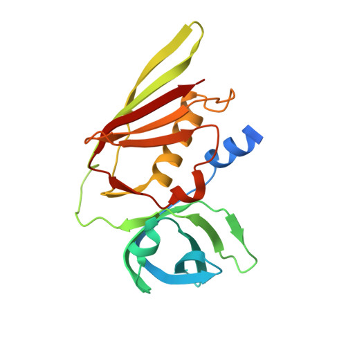
Crystal structures of the staphylococcal toxin SSL5 in complex with sialyl Lewis X reveal a conserved binding site that shares common features with viral and bacterial sialic acid binding proteins

Baker, H.M.Basu, I.Chung, M.C.Caradoc-Davies, T.Fraser, J.D.Baker, E.N.

(2007) J Mol Biol 374: 1298-1308

  • DOI: https://doi.org/10.1016/j.jmb.2007.09.091
  • Primary Citation of Related Structures:  
    2R61, 2Z8L

  • PubMed Abstract: 

    Staphylococcus aureus is a significant human pathogen. Among its large repertoire of secreted toxins is a group of staphylococcal superantigen-like proteins (SSLs). These are homologous to superantigens but do not have the same activity. SSL5 is shown here to bind to human granulocytes and to the cell surface receptors for human IgA (Fc alphaRI) and P-selectin [P-selectin glycoprotein ligand-1 (PSGL-1)] in a sialic acid (Sia)-dependent manner. Co-crystallization of SSL5 with the tetrasaccharide sialyl Lewis X (sLe(X)), a key determinant of PSGL-1 binding to P-selectin, led to crystal structures of the SSL5-sLe(X) complex at resolutions of 1.65 and 2.75 A for crystals at two pH values. In both structures, sLe(X) bound to a specific site on the surface of the C-terminal domain of SSL5 in a conformation identical with that bound by P-selectin. Conservation of the key carbohydrate binding residues indicates that this ability to bind human glycans is shared by a substantial subgroup of the SSLs, including SSL2, SSL3, SSL4, SSL5, SSL6, and SSL11. This indicates that the ability to target human glycans is an important property of this group of toxins. Structural comparisons also showed that the Sia binding site in SSL5 contains a substructure that is shared by other Sia binding proteins from bacteria as well as viruses and represents a common binding motif.


  • Organizational Affiliation

    Maurice Wilkins Centre for Molecular Biodiscovery, University of Auckland, Auckland, New Zealand.


Macromolecules
Find similar proteins by:  (by identity cutoff)  |  3D Structure
Entity ID: 1
MoleculeChains Sequence LengthOrganismDetailsImage
Exotoxin 3208Staphylococcus aureusMutation(s): 0 
Gene Names: ssl5
UniProt
Find proteins for Q9ZFS6 (Staphylococcus aureus)
Explore Q9ZFS6 
Go to UniProtKB:  Q9ZFS6
Entity Groups  
Sequence Clusters30% Identity50% Identity70% Identity90% Identity95% Identity100% Identity
UniProt GroupQ9ZFS6
Sequence Annotations
Expand
  • Reference Sequence
Oligosaccharides

Help

Entity ID: 2
MoleculeChains Length2D Diagram Glycosylation3D Interactions
N-acetyl-alpha-neuraminic acid-(2-3)-beta-D-galactopyranose-(1-4)-[alpha-L-fucopyranose-(1-3)]2-acetamido-2-deoxy-beta-D-glucopyranose
B
4N/A
Glycosylation Resources
GlyTouCan:  G00054MO
GlyCosmos:  G00054MO
GlyGen:  G00054MO
Biologically Interesting Molecules (External Reference) 1 Unique
Experimental Data & Validation

Experimental Data

  • Method: X-RAY DIFFRACTION
  • Resolution: 1.65 Å
  • R-Value Free: 0.218 
  • R-Value Work: 0.181 
  • R-Value Observed: 0.183 
  • Space Group: P 1 21 1
Unit Cell:
Length ( Å )Angle ( ˚ )
a = 37.625α = 90
b = 78.237β = 127.02
c = 55.276γ = 90
Software Package:
Software NamePurpose
MOSFLMdata reduction
PHASERphasing
REFMACrefinement
PDB_EXTRACTdata extraction
HKL-2000data collection
SCALAdata scaling

Structure Validation

View Full Validation Report


Entry History 

Deposition Data

Revision History  (Full details and data files)

  • Version 1.0: 2007-11-27
    Type: Initial release
  • Version 1.1: 2011-07-13
    Changes: Advisory, Version format compliance
  • Version 2.0: 2020-07-29
    Type: Remediation
    Reason: Carbohydrate remediation
    Changes: Atomic model, Data collection, Database references, Derived calculations, Structure summary
  • Version 2.1: 2023-11-01
    Changes: Data collection, Database references, Refinement description, Structure summary