Experimental Data Snapshot

  • Method: X-RAY DIFFRACTION
  • Resolution: 2.20 Å
  • R-Value Free: 
    0.222 (Depositor), 0.225 (DCC) 
  • R-Value Work: 
    0.190 (Depositor), 0.197 (DCC) 
  • R-Value Observed: 
    0.192 (Depositor) 

wwPDB Validation 3D Report Full Report

Validation slider image for 2YAD

This is version 1.3 of the entry. See complete history

Literature

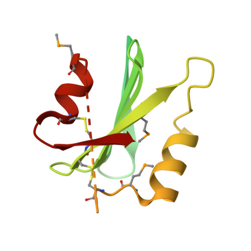
High Resolution Structure of a Bricos Domain and its Implications for Anti-Amyloid Chaperone Activity on Lung Surgactant Protein C.

Willander, H.Askarieh, G.Landreh, M.Westermark, P.Nordling, K.Keranen, H.Hermansson, E.Hamvas, A.Nogee, L.M.Bergman, T.Saenz, A.Casals, C.Aqvist, J.Jornvall, H.Berglund, H.Presto, J.Knight, S.D.Johansson, J.

(2012) Proc Natl Acad Sci U S A 109: 2325

  • DOI: https://doi.org/10.1073/pnas.1114740109
  • Primary Citation Related Structures: 
    2YAD

  • PubMed Abstract: 

    BRICHOS domains are encoded in > 30 human genes, which are associated with cancer, neurodegeneration, and interstitial lung disease (ILD). The BRICHOS domain from lung surfactant protein C proprotein (proSP-C) is required for membrane insertion of SP-C and has anti-amyloid activity in vitro. Here, we report the 2.1 Å crystal structure of the human proSP-C BRICHOS domain, which, together with molecular dynamics simulations and hydrogen-deuterium exchange mass spectrometry, reveals how BRICHOS domains may mediate chaperone activity. Observation of amyloid deposits composed of mature SP-C in lung tissue samples from ILD patients with mutations in the BRICHOS domain or in its peptide-binding linker region supports the in vivo relevance of the proposed mechanism. The results indicate that ILD mutations interfering with proSP-C BRICHOS activity cause amyloid disease secondary to intramolecular chaperone malfunction.


  • Organizational Affiliation
    • Department of Anatomy, Physiology, and Biochemistry, Swedish University of Agricultural Sciences, S-751 24 Uppsala, Sweden.

Macromolecule Content 

  • Total Structure Weight: 112.25 kDa 
  • Atom Count: 3,697 
  • Modeled Residue Count: 471 
  • Deposited Residue Count: 1,026 
  • Unique protein chains: 1

Macromolecules

Find similar proteins by:|  3D Structure
Entity ID: 1
MoleculeChains  Sequence LengthOrganismDetailsImage
SURFACTANT PROTEIN C BRICHOS DOMAIN
A, B, C, D, E
A, B, C, D, E, F
171Homo sapiensMutation(s): 0 
UniProt & NIH Common Fund Data Resources
Find proteins for P11686 (Homo sapiens)
Explore P11686 
Go to UniProtKB:  P11686
PHAROS:  P11686
GTEx:  ENSG00000168484 
Entity Groups
Sequence Clusters30% Identity50% Identity70% Identity90% Identity95% Identity100% Identity
UniProt GroupP11686
Sequence Annotations
Expand
Reference Sequence

Small Molecules

Modified Residues  1 Unique
IDChains TypeFormula2D DiagramParent
MSE
Query on MSE
A, B, C, D, E
A, B, C, D, E, F
L-PEPTIDE LINKINGC5 H11 N O2 SeMET

Experimental Data & Validation

Experimental Data

  • Method: X-RAY DIFFRACTION
  • Resolution: 2.20 Å
  • R-Value Free:  0.222 (Depositor), 0.225 (DCC) 
  • R-Value Work:  0.190 (Depositor), 0.197 (DCC) 
  • R-Value Observed: 0.192 (Depositor) 
Space Group: C 1 2 1
Unit Cell:
Length ( Å )Angle ( ˚ )
a = 132.046α = 90
b = 39.329β = 99.63
c = 114.756γ = 90
Software Package:
Software NamePurpose
BUSTERrefinement

Structure Validation

View Full Validation Report


Entry History 

Revision History  (Full details and data files)

  • Version 1.0: 2012-02-15
    Type: Initial release
  • Version 1.1: 2012-02-29
    Changes: Other
  • Version 1.2: 2014-08-13
    Changes: Refinement description
  • Version 1.3: 2024-10-23
    Changes: Data collection, Database references, Derived calculations, Other, Structure summary