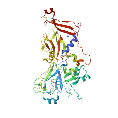
2Y8R

Crystal structure of apo AMA1 mutant (Tyr230Ala) from Toxoplasma gondii


Experimental Data Snapshot

  • Method: X-RAY DIFFRACTION
  • Resolution: 2.45 Å
  • R-Value Free: 0.305 
  • R-Value Work: 0.212 
  • R-Value Observed: 0.216 

wwPDB Validation   3D Report Full Report


Ligand Structure Quality Assessment 


This is version 1.2 of the entry. See complete history


Literature

Host Cell Invasion by Apicomplexan Parasites: Insights from the Co-Structure of Ama1 with a Ron2 Peptide

Tonkin, M.L.Roques, M.Lamarque, M.H.Pugniere, M.Douguet, D.Crawford, J.Lebrun, M.Boulanger, M.J.

(2011) Science 333: 463

  • DOI: https://doi.org/10.1126/science.1204988
  • Primary Citation of Related Structures:  
    2Y8R, 2Y8S, 2Y8T

  • PubMed Abstract: 

    Apicomplexan parasites such as Toxoplasma gondii and Plasmodium species actively invade host cells through a moving junction (MJ) complex assembled at the parasite-host cell interface. MJ assembly is initiated by injection of parasite rhoptry neck proteins (RONs) into the host cell, where RON2 spans the membrane and functions as a receptor for apical membrane antigen 1 (AMA1) on the parasite. We have determined the structure of TgAMA1 complexed with a RON2 peptide at 1.95 angstrom resolution. A stepwise assembly mechanism results in an extensive buried surface area, enabling the MJ complex to resist the mechanical forces encountered during host cell invasion. Besides providing insights into host cell invasion by apicomplexan parasites, the structure offers a basis for designing therapeutics targeting these global pathogens.


  • Organizational Affiliation

    Department of Biochemistry and Microbiology, University of Victoria, Victoria, British Columbia V8W 3P6, Canada.


Macromolecules
Find similar proteins by:  (by identity cutoff)  |  3D Structure
Entity ID: 1
MoleculeChains Sequence LengthOrganismDetailsImage
APICAL MEMBRANE ANTIGEN, PUTATIVEA,
B,
C [auth D],
D [auth E]
432Toxoplasma gondiiMutation(s): 1 
UniProt
Find proteins for B6KAM0 (Toxoplasma gondii (strain ATCC 50861 / VEG))
Explore B6KAM0 
Go to UniProtKB:  B6KAM0
Entity Groups  
Sequence Clusters30% Identity50% Identity70% Identity90% Identity95% Identity100% Identity
UniProt GroupB6KAM0
Glycosylation
Glycosylation Sites: 1
Sequence Annotations
Expand
  • Reference Sequence
Experimental Data & Validation

Experimental Data

  • Method: X-RAY DIFFRACTION
  • Resolution: 2.45 Å
  • R-Value Free: 0.305 
  • R-Value Work: 0.212 
  • R-Value Observed: 0.216 
  • Space Group: P 1
Unit Cell:
Length ( Å )Angle ( ˚ )
a = 67.52α = 71.89
b = 76.09β = 73.37
c = 88.78γ = 73.5
Software Package:
Software NamePurpose
REFMACrefinement
PHASERphasing

Structure Validation

View Full Validation Report


Ligand Structure Quality Assessment 


Entry History 

Deposition Data

Revision History  (Full details and data files)

  • Version 1.0: 2011-08-03
    Type: Initial release
  • Version 1.1: 2019-04-03
    Changes: Data collection, Derived calculations, Other, Source and taxonomy
  • Version 1.2: 2020-07-29
    Type: Remediation
    Reason: Carbohydrate remediation
    Changes: Data collection, Derived calculations, Other, Structure summary