2Y35 | pdb_00002y35

Crystal structure of Xrn1-substrate complex


Experimental Data Snapshot

  • Method: X-RAY DIFFRACTION
  • Resolution: 3.20 Å
  • R-Value Free: 
    0.271 (Depositor), 0.271 (DCC) 
  • R-Value Work: 
    0.225 (Depositor), 0.223 (DCC) 
  • R-Value Observed: 
    0.227 (Depositor) 

wwPDB Validation 3D Report Full Report

Validation slider image for 2Y35

This is version 1.2 of the entry. See complete history

Literature

Coupled 5' Nucleotide Recognition and Processivity in Xrn1-Mediated Mrna Decay.

Jinek, M.Coyle, S.M.Doudna, J.A.

(2011) Mol Cell 41: 600

  • DOI: https://doi.org/10.1016/j.molcel.2011.02.004
  • Primary Citation Related Structures: 
    2Y35

  • PubMed Abstract: 

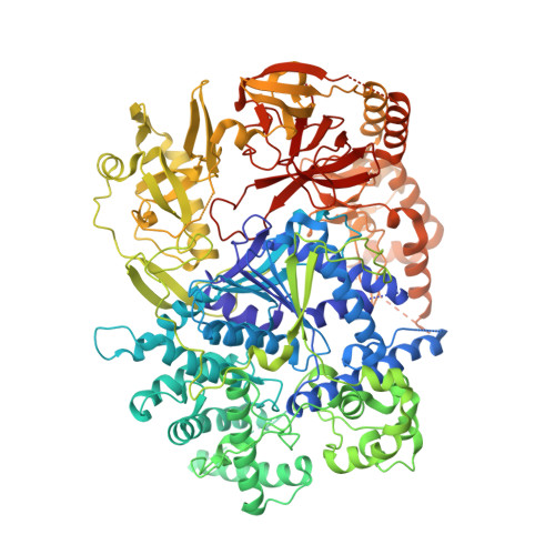
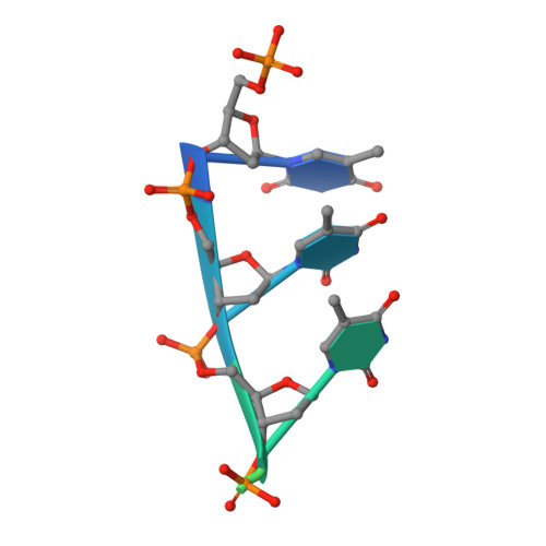
    Messenger RNA decay plays a central role in the regulation and surveillance of eukaryotic gene expression. The conserved multidomain exoribonuclease Xrn1 targets cytoplasmic RNA substrates marked by a 5' monophosphate for processive 5'-to-3' degradation by an unknown mechanism. Here, we report the crystal structure of an Xrn1-substrate complex. The single-stranded substrate is held in place by stacking of the 5'-terminal trinucleotide between aromatic side chains while a highly basic pocket specifically recognizes the 5' phosphate. Mutations of residues involved in binding the 5'-terminal nucleotide impair Xrn1 processivity. The substrate recognition mechanism allows Xrn1 to couple processive hydrolysis to duplex melting in RNA substrates with sufficiently long single-stranded 5' overhangs. The Xrn1-substrate complex structure thus rationalizes the exclusive specificity of Xrn1 for 5'-monophosphorylated substrates, ensuring fidelity of mRNA turnover, and posits a model for translocation-coupled unwinding of structured RNA substrates.


  • Organizational Affiliation
    • Department of Molecular and Cell Biology, University of California, Berkeley, Berkeley, CA 94720, USA.

Macromolecule Content 

  • Total Structure Weight: 135.72 kDa 
  • Atom Count: 8,187 
  • Modeled Residue Count: 1,030 
  • Deposited Residue Count: 1,151 
  • Unique protein chains: 1
  • Unique nucleic acid chains: 1

Macromolecules


Find similar proteins by:|  3D Structure
Entity ID: 1
MoleculeChains  Sequence LengthOrganismDetailsImage
LD22664P1,140Drosophila melanogasterMutation(s): 1 
EC: 3.1.13
UniProt
Find proteins for E1JJR3 (Drosophila melanogaster)
Explore E1JJR3 
Go to UniProtKB:  E1JJR3
Entity Groups
Sequence Clusters30% Identity50% Identity70% Identity90% Identity95% Identity100% Identity
UniProt GroupE1JJR3
Sequence Annotations
Expand
Reference Sequence
Find similar nucleic acids by:  Sequence
Entity ID: 2
MoleculeChains LengthOrganismImage
DT11 (5'-D(*TP*TP*TP*TP*TP*TP*TP*TP*TP*TP*TP)-3'11synthetic construct
Sequence Annotations
Expand
Reference Sequence

Small Molecules

Ligands 1 Unique
IDChains Name / Formula / InChI Key2D Diagram3D Interactions
MG

Query on MG


Download:Ideal Coordinates CCD File
C [auth A]MAGNESIUM ION
Mg
JLVVSXFLKOJNIY-UHFFFAOYSA-N

Experimental Data & Validation

Experimental Data

  • Method: X-RAY DIFFRACTION
  • Resolution: 3.20 Å
  • R-Value Free:  0.271 (Depositor), 0.271 (DCC) 
  • R-Value Work:  0.225 (Depositor), 0.223 (DCC) 
  • R-Value Observed: 0.227 (Depositor) 
Space Group: P 42 21 2
Unit Cell:
Length ( Å )Angle ( ˚ )
a = 149.99α = 90
b = 149.99β = 90
c = 154.86γ = 90
Software Package:
Software NamePurpose
XDSdata reduction
XSCALEdata scaling
PHENIXphasing
autoSHARPphasing
PHENIXrefinement

Structure Validation

View Full Validation Report


Entry History 

Deposition Data

Revision History  (Full details and data files)

  • Version 1.0: 2011-03-16
    Type: Initial release
  • Version 1.1: 2011-08-03
    Changes: Derived calculations, Version format compliance
  • Version 1.2: 2024-05-08
    Changes: Data collection, Database references, Derived calculations, Other