2XTS

Crystal Structure of the Sulfane Dehydrogenase SoxCD from Paracoccus pantotrophus


Experimental Data Snapshot

  • Method: X-RAY DIFFRACTION
  • Resolution: 1.33 Å
  • R-Value Free: 0.111 
  • R-Value Work: 0.107 
  • R-Value Observed: 0.107 

wwPDB Validation   3D Report Full Report


Ligand Structure Quality Assessment 


This is version 1.2 of the entry. See complete history


Literature

Structural Basis for the Oxidation of Protein-Bound Sulfur by the Sulfur Cycle Molybdohemo-Enzyme Sulfane Dehydrogenase Soxcd.

Zander, U.Faust, A.Klink, B.U.De Sanctis, D.Panjikar, S.Quentmeier, A.Bardischewsky, F.Friedrich, C.G.Scheidig, A.J.

(2011) J Biol Chem 286: 8349

  • DOI: https://doi.org/10.1074/jbc.M110.193631
  • Primary Citation of Related Structures:  
    2XTS

  • PubMed Abstract: 

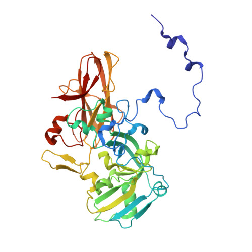
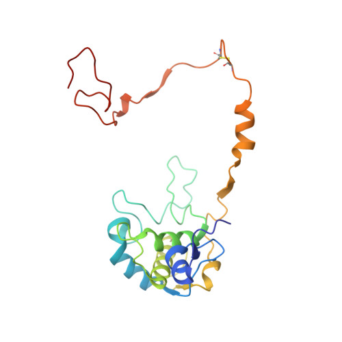
    The sulfur cycle enzyme sulfane dehydrogenase SoxCD is an essential component of the sulfur oxidation (Sox) enzyme system of Paracoccus pantotrophus. SoxCD catalyzes a six-electron oxidation reaction within the Sox cycle. SoxCD is an α(2)β(2) heterotetrameric complex of the molybdenum cofactor-containing SoxC protein and the diheme c-type cytochrome SoxD with the heme domains D(1) and D(2). SoxCD(1) misses the heme-2 domain D(2) and is catalytically as active as SoxCD. The crystal structure of SoxCD(1) was solved at 1.33 Å. The substrate of SoxCD is the outer (sulfane) sulfur of Cys-110-persulfide located at the C-terminal peptide swinging arm of SoxY of the SoxYZ carrier complex. The SoxCD(1) substrate funnel toward the molybdopterin is narrow and partially shielded by side-chain residues of SoxD(1). For access of the sulfane-sulfur of SoxY-Cys-110 persulfide we propose that (i) the blockage by SoxD-Arg-98 is opened via interaction with the C terminus of SoxY and (ii) the C-terminal peptide VTIGGCGG of SoxY provides interactions with the entrance path such that the cysteine-bound persulfide is optimally positioned near the molybdenum atom. The subsequent oxidation reactions of the sulfane-sulfur are initiated by the nucleophilic attack of the persulfide anion on the molybdenum atom that is, in turn, reduced. The close proximity of heme-1 to the molybdopterin allows easy acceptance of the electrons. Because SoxYZ, SoxXA, and SoxB are already structurally characterized, with SoxCD(1) the structures of all key enzymes of the Sox cycle are known with atomic resolution.


  • Organizational Affiliation

    From the Department of Structural Biology, Zoological Institute, Christian-Albrechts-University Kiel, 24118 Kiel, Germany,; the Department of Biophysics-Structural Biology, Saarland University, 66421 Homburg, Germany.


Macromolecules
Find similar proteins by:  (by identity cutoff)  |  3D Structure
Entity ID: 1
MoleculeChains Sequence LengthOrganismDetailsImage
SULFITE DEHYDROGENASE
A, C
390Paracoccus pantotrophusMutation(s): 0 
EC: 1.8.2.1
UniProt
Find proteins for P72178 (Paracoccus denitrificans)
Explore P72178 
Go to UniProtKB:  P72178
Entity Groups  
Sequence Clusters30% Identity50% Identity70% Identity90% Identity95% Identity100% Identity
UniProt GroupP72178
Sequence Annotations
Expand
  • Reference Sequence
Find similar proteins by:  (by identity cutoff)  |  3D Structure
Entity ID: 2
MoleculeChains Sequence LengthOrganismDetailsImage
CYTOCHROME
B, D
205Paracoccus pantotrophusMutation(s): 0 
UniProt
Find proteins for O07819 (Paracoccus denitrificans)
Explore O07819 
Go to UniProtKB:  O07819
Entity Groups  
Sequence Clusters30% Identity50% Identity70% Identity90% Identity95% Identity100% Identity
UniProt GroupO07819
Sequence Annotations
Expand
  • Reference Sequence
Small Molecules
Ligands 6 Unique
IDChains Name / Formula / InChI Key2D Diagram3D Interactions
HEC
Query on HEC

Download Ideal Coordinates CCD File 
K [auth B],
U [auth D]
HEME C
C34 H34 Fe N4 O4
HXQIYSLZKNYNMH-LJNAALQVSA-N
MTE
Query on MTE

Download Ideal Coordinates CCD File 
E [auth A],
P [auth C]
PHOSPHONIC ACIDMONO-(2-AMINO-5,6-DIMERCAPTO-4-OXO-3,7,8A,9,10,10A-HEXAHYDRO-4H-8-OXA-1,3,9,10-TETRAAZA-ANTHRACEN-7-YLMETHYL)ESTER
C10 H14 N5 O6 P S2
HPEUEJRPDGMIMY-IFQPEPLCSA-N
2MO
Query on 2MO

Download Ideal Coordinates CCD File 
F [auth A],
Q [auth C]
MOLYBDENUM (IV)OXIDE
Mo O2
QXYJCZRRLLQGCR-UHFFFAOYSA-N
GOL
Query on GOL

Download Ideal Coordinates CCD File 
AA [auth D]
H [auth A]
I [auth A]
J [auth A]
N [auth B]
AA [auth D],
H [auth A],
I [auth A],
J [auth A],
N [auth B],
O [auth B],
S [auth C],
T [auth C],
Z [auth D]
GLYCEROL
C3 H8 O3
PEDCQBHIVMGVHV-UHFFFAOYSA-N
CO
Query on CO

Download Ideal Coordinates CCD File 
G [auth A],
R [auth C]
COBALT (II) ION
Co
XLJKHNWPARRRJB-UHFFFAOYSA-N
CA
Query on CA

Download Ideal Coordinates CCD File 
L [auth B]
M [auth B]
V [auth D]
W [auth D]
X [auth D]
L [auth B],
M [auth B],
V [auth D],
W [auth D],
X [auth D],
Y [auth D]
CALCIUM ION
Ca
BHPQYMZQTOCNFJ-UHFFFAOYSA-N
Experimental Data & Validation

Experimental Data

  • Method: X-RAY DIFFRACTION
  • Resolution: 1.33 Å
  • R-Value Free: 0.111 
  • R-Value Work: 0.107 
  • R-Value Observed: 0.107 
  • Space Group: P 31
Unit Cell:
Length ( Å )Angle ( ˚ )
a = 122.97α = 90
b = 122.97β = 90
c = 76.39γ = 120
Software Package:
Software NamePurpose
REFMACrefinement
XDSdata reduction
SCALAdata scaling
Auto-Rickshawphasing

Structure Validation

View Full Validation Report


Ligand Structure Quality Assessment 


Entry History 

Deposition Data

Revision History  (Full details and data files)

  • Version 1.0: 2010-12-08
    Type: Initial release
  • Version 1.1: 2011-05-08
    Changes: Version format compliance
  • Version 1.2: 2011-07-13
    Changes: Version format compliance