A Hot Oxidant, 3-No(2)Y(122) Radical, Unmasks Conformational Gating in Ribonucleotide Reductase.
Yokoyama, K., Uhlin, U., Stubbe, J.(2010) J Am Chem Soc 132: 15368
- PubMed: 20929229 Search on PubMedSearch on PubMed Central
- DOI: https://doi.org/10.1021/ja1069344
- Primary Citation Related Structures:
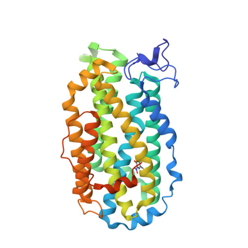
2XOF - PubMed Abstract:
Escherichia coli ribonucleotide reductase is an α2β2 complex that catalyzes the conversion of nucleotides to deoxynucleotides and requires a diferric-tyrosyl radical (Y(•)) cofactor to initiate catalysis. The initiation process requires long-range proton-coupled electron transfer (PCET) over 35 Å between the two subunits by a specific pathway (Y(122)(•)→W(48)→Y(356) within β to Y(731)→Y(730)→C(439) within α). The rate-limiting step in nucleotide reduction is the conformational gating of the PCET process, which masks the chemistry of radical propagation. 3-Nitrotyrosine (NO(2)Y) has recently been incorporated site-specifically in place of Y(122) in β2. The protein as isolated contained a diferric cluster but no nitrotyrosyl radical (NO(2)Y(•)) and was inactive. In the present paper we show that incubation of apo-Y(122)NO(2)Y-β2 with Fe(2+) and O(2) generates a diferric-NO(2)Y(•) that has a half-life of 40 s at 25 °C. Sequential mixing experiments, in which the cofactor is assembled to 1.2 NO(2)Y(•)/β2 and then mixed with α2, CDP, and ATP, have been analyzed by stopped-flow absorption spectroscopy, rapid freeze quench EPR spectroscopy, and rapid chemical quench methods. These studies have, for the first time, unmasked the conformational gating. They reveal that the NO(2)Y(•) is reduced to the nitrotyrosinate with biphasic kinetics (283 and 67 s(-1)), that dCDP is produced at 107 s(-1), and that a new Y(•) is produced at 97 s(-1). Studies with pathway mutants suggest that the new Y(•) is predominantly located at 356 in β2. In consideration of these data and the crystal structure of Y(122)NO(2)Y-β2, a mechanism for PCET uncoupling in NO(2)Y(•)-RNR is proposed.
- Department of Chemistry, Massachusetts Institute of Technology, 77 Massachusetts Avenue, Cambridge, Massachusetts 02139-4307, United States.
Organizational Affiliation: