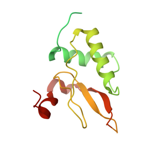
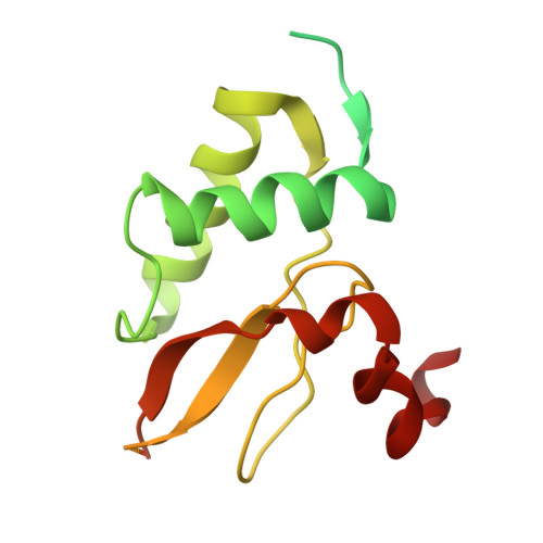
A Novel Dimer-Tetramer Transition Captured by the Crystal Structure of the HIV-1 Nef.
Singh, P., Yadav, G.P., Gupta, S., Tripathi, A.K., Ramachandran, R., Tripathi, R.K.(2011) PLoS One 6: 26629
- PubMed: 22073177 Search on PubMedSearch on PubMed Central
- DOI: https://doi.org/10.1371/journal.pone.0026629
- Primary Citation Related Structures:
2XI1 - PubMed Abstract:
HIV-1 Nef modulates disease progression through interactions with over 30 host proteins. Individual chains fold into membrane-interacting N-terminal and C-terminal core (Nef(core)) domains respectively. Nef exists as small oligomers near membranes and associates into higher oligomers such as tetramers or hexadecamers in the cytoplasm. Earlier structures of the Nef(core) in apo and complexed forms with the Fyn-kinase SH3 domain revealed dimeric association details and the role of the conserved PXXP recognition motif (residues 72-78) of Nef in SH3-domain interactions. The crystal structure of the tetrameric Nef reported here corresponds to the elusive cytoplasmic stage. Comparative analyses show that subunits of Nef(core) dimers (open conformation) swing out with a relative displacement of ~22 Å and rotation of ~174° to form the 'closed' tetrameric structure. The changes to the association are around Asp125, a conserved residue important for viral replication and the important XR motif (residues 107-108). The tetramer associates through C4 symmetry instead of the 222 symmetry expected when two dimers associate together. This novel dimer-tetramer transition agrees with earlier solution studies including small angle X-ray scattering, analytical ultracentrifugation, dynamic laser light scattering and our glutaraldehyde cross-linking experiments. Comparisons with the Nef(core)--Fyn-SH3 domain complexes reveal that the PXXP motif that interacts with the SH3-domain in the dimeric form is sterically occluded in the tetramer. However the 151-180 loop that is distal to the PXXP motif and contains several protein interaction motifs remains accessible. The results suggest how changes to the oligomeric state of Nef can help it distinguish between protein partners.
- Toxicology Division, Central Drug Research Institute, Council of Scientific & Industrial Research, Chattar Manzil, Mahatma Gandhi Marg, Lucknow, Uttar Pradesh, India.
Organizational Affiliation: