Structure of a Family 3B' Carbohydrate-Binding Module from the Cel9V Glycoside Hydrolase from Clostridium Thermocellum: Structural Diversity and Implications for Carbohydrate Binding
Petkun, S., Jindou, S., Shimon, L.J.W., Bayer, E.A., Lamed, R., Frolow, F.(2010) Acta Crystallogr D Biol Crystallogr 66: 33
- PubMed: 20057047 Search on PubMed
- DOI: https://doi.org/10.1107/S0907444909043030
- Primary Citation Related Structures:
2WNX, 2WO4, 2WOB - PubMed Abstract:
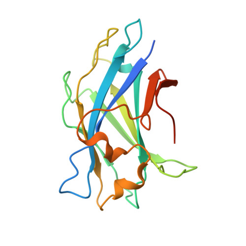
Family 3 carbohydrate-binding modules (CBM3s) are associated with both cellulosomal scaffoldins and family 9 glycoside hydrolases (GH9s), which are multi-modular enzymes that act on cellulosic substrates. CBM3s bind cellulose. X-ray crystal structures of these modules have established an accepted cellulose-binding mechanism based on stacking interactions between the sugar rings of cellulose and a planar array of aromatic residues located on the CBM3 surface. These planar-strip residues are generally highly conserved, although some CBM3 sequences lack one or more of these residues. In particular, CBM3b' from Clostridium thermocellum Cel9V exhibits such sequence changes and fails to bind cellulosic substrates. A crystallographic investigation of CBM3b' has been initiated in order to understand the structural reason(s) for this inability. CBM3b' crystallized in space group C222(1) (diffraction was obtained to 2.0 A resolution in-house) with three independent molecules in the asymmetric unit and in space group P4(1)2(1)2 (diffraction was obtained to 1.79 A resolution in-house and to 1.30 A resolution at a synchrotron) with one molecule in the asymmetric unit. The molecular structure of Cel9V CBM3b' revealed that in addition to the loss of several cellulose-binding residues in the planar strip, changes in the backbone create a surface 'hump' which could interfere with the formation of cellulose-protein surface interactions and thus prevent binding to crystalline cellulose.
- Department of Molecular Microbiology and Biotechnology, Tel Aviv University, Tel Aviv 69978, Israel.
Organizational Affiliation: