Crystal Structures and Enzyme Mechanism of a Dual Fucose Mutarotase/Ribose Pyranase
Lee, K.-H., Ryu, K.-S., Kim, M.-S., Suh, H.-Y., Ku, B., Song, Y.-L., Ko, S., Lee, W., Oh, B.-H.(2009) J Mol Biology 391: 178
- PubMed: 19524593
- DOI: https://doi.org/10.1016/j.jmb.2009.06.022
- Primary Citation of Related Structures:
2WCU, 2WCV - PubMed Abstract:
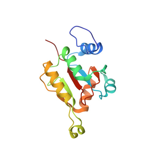
Escherichia coli FucU (Fucose Unknown) is a dual fucose mutarotase and ribose pyranase, which shares 44% sequence identity with its human counterpart. Herein, we report the structures of E. coli FucU and mouse FucU bound to L-fucose and delineate the catalytic mechanisms underlying the interconversion between stereoisomers of fucose and ribose. E. coli FucU forms a decameric toroid with each active site formed by two adjacent subunits. While one subunit provides most of the fucose-interacting residues including a catalytic tyrosine residue, the other subunit provides a catalytic His-Asp dyad. This active-site feature is critical not only for the mutarotase activity toward L-fucose but also for the pyranase activity toward D-ribose. Structural and biochemical analyses pointed that mouse FucU assembles into four different oligomeric forms, among which the smallest homodimeric form is most abundant and would be the predominant species under physiological conditions. This homodimer has two fucose-binding sites that are devoid of the His-Asp dyad and catalytically inactive, indicating that the mutarotase and the pyranase activities appear dispensable in vertebrates. The defective assembly of the mouse FucU homodimer into the decameric form is due to an insertion of two residues at the N-terminal extreme, which is a common aspect of all the known vertebrate FucU proteins. Therefore, vertebrate FucU appears to serve for as yet unknown function through the quaternary structural alteration.
- Center for Biomolecular Recognition, Division of Molecular and Life Sciences, Pohang University of Science and Technology, Pohang, Kyungbuk, Korea.
Organizational Affiliation:


