2W52 | pdb_00002w52

2 beta-glucans (6-O-glucosyl-laminaritriose) in both donor and acceptor sites of GH16 Laminarinase 16A from Phanerochaete chrysosporium.


Experimental Data Snapshot

  • Method: X-RAY DIFFRACTION
  • Resolution: 1.56 Å
  • R-Value Free: 
    0.171 (Depositor), 0.171 (DCC) 
  • R-Value Work: 
    0.151 (Depositor), 0.149 (DCC) 
  • R-Value Observed: 
    0.152 (Depositor) 

wwPDB Validation 3D Report Full Report

Validation slider image for 2W52

This is version 2.1 of the entry. See complete history. 

Literature

X-Ray Crystal Structures of Phanerochaete Chrysosporium Laminarinase 16A in Complex with Products from Lichenin and Laminarin Hydrolysis

Vasur, J., Kawai, R., Andersson, E., Igarashi, K., Sandgren, M., Samejima, M., Stahlberg, J.

(2009) FEBS J 276: 3858

  • DOI: https://doi.org/10.1111/j.1742-4658.2009.07099.x
  • Primary Citation Related Structures: 
    2W39, 2W52

  • PubMed Abstract: 

    The 1,3(4)-beta-D-glucanases of glycoside hydrolase family 16 provide useful examples of versatile yet specific protein-carbohydrate interactions. In the present study, we report the X-ray structures of the 1,3(4)-beta-D-glucanase Phanerochaete chrysosporium Laminarinase 16A in complex with beta-glucan products from laminarin (1.6 A) and lichenin (1.1 A) hydrolysis. The G6G3G3G glucan, in complex with the enzyme, showed a beta-1,6 branch in the acceptor site. The G4G3G ligand-protein complex showed that there was no room for a beta-1,6 branch in the -1 or -2 subsites; furthermore, the distorted residue in the -1 subsite and the glucose in the -2 subsite required a beta-1,3 bond between them. These are the first X-ray crystal structures of any 1,3(4)-beta-D-glucanase in complex with glucan products. They provide details of both substrate and product binding in support of earlier enzymatic evidence.


  • Organizational Affiliation: 
    • Department of Molecular Biology, University of Agricultural Sciences, Uppsala, Sweden.

Macromolecule Content 

  • Total Structure Weight: 34.33 kDa 
  • Atom Count: 2,871 
  • Modeled Residue Count: 298 
  • Deposited Residue Count: 298 
  • Unique protein chains: 1

Macromolecules

Find similar proteins by:|  3D Structure
Entity ID: 1
MoleculeChains  Sequence LengthOrganismDetailsImage
PUTATIVE LAMINARINASE298Phanerodontia chrysosporiumMutation(s): 0 
EC: 3.2.1.6
UniProt
Find proteins for Q874E3 (Phanerodontia chrysosporium)
Explore Q874E3 
Go to UniProtKB:  Q874E3
Entity Groups
Sequence Clusters30% Identity50% Identity70% Identity90% Identity95% Identity100% Identity
UniProt GroupQ874E3
Glycosylation
Glycosylation Sites: 1
Sequence Annotations
Expand
Reference Sequence

Oligosaccharides

Help  
Entity ID: 2
MoleculeChains Length2D Diagram GlycosylationD Interactions
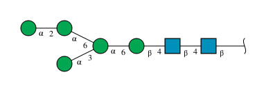
alpha-D-mannopyranose-(1-2)-alpha-D-mannopyranose-(1-6)-[alpha-D-mannopyranose-(1-3)]alpha-D-mannopyranose-(1-6)-beta-D-mannopyranose-(1-4)-2-acetamido-2-deoxy-beta-D-glucopyranose-(1-4)-2-acetamido-2-deoxy-beta-D-glucopyranose
B
7N-Glycosylation
Glycosylation Resources
GlyTouCan: G94559IJ
GlyCosmos: G94559IJ
GlyGen: G94559IJ
Entity ID: 3
MoleculeChains Length2D Diagram GlycosylationD Interactions
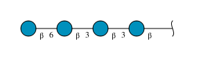
beta-D-glucopyranose-(1-6)-beta-D-glucopyranose-(1-3)-beta-D-glucopyranose-(1-3)-beta-D-glucopyranose
C
4N/A
Glycosylation Resources
GlyTouCan: G19503WF
GlyCosmos: G19503WF
GlyGen: G19503WF
Entity ID: 4
MoleculeChains Length2D Diagram GlycosylationD Interactions
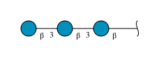
beta-D-glucopyranose-(1-3)-beta-D-glucopyranose-(1-3)-beta-D-glucopyranose
D
3N/A
Glycosylation Resources
GlyTouCan: G00024MO
GlyCosmos: G00024MO
GlyGen: G00024MO

Experimental Data & Validation

Experimental Data

  • Method: X-RAY DIFFRACTION
  • Resolution: 1.56 Å
  • R-Value Free:  0.171 (Depositor), 0.171 (DCC) 
  • R-Value Work:  0.151 (Depositor), 0.149 (DCC) 
  • R-Value Observed: 0.152 (Depositor) 
Space Group: P 21 21 21
Unit Cell:
Length ( Å )Angle ( ˚ )
a = 38.056α = 90
b = 46.69β = 90
c = 152.871γ = 90
Software Package:
Software NamePurpose
REFMACrefinement
MOSFLMdata reduction
SCALAdata scaling
REFMACphasing

Structure Validation

View Full Validation Report


Entry History 

Deposition Data

Revision History  (Full details and data files)

  • Version 1.0: 2009-07-21
    Type: Initial release
  • Version 1.1: 2012-04-04
    Changes: Database references, Derived calculations, Non-polymer description, Other, Structure summary, Version format compliance
  • Version 1.2: 2018-01-17
    Changes: Data collection
  • Version 2.0: 2020-07-29
    Type: Remediation
    Reason: Carbohydrate remediation
    Changes: Atomic model, Data collection, Derived calculations, Other, Structure summary
  • Version 2.1: 2024-10-23
    Changes: Data collection, Database references, Structure summary