2W16

Structures of FpvA bound to heterologous pyoverdines


Experimental Data Snapshot

  • Method: X-RAY DIFFRACTION
  • Resolution: 2.71 Å
  • R-Value Free: 0.253 
  • R-Value Work: 0.217 
  • R-Value Observed: 0.219 

wwPDB Validation   3D Report Full Report


Ligand Structure Quality Assessment 


This is version 1.6 of the entry. See complete history


Literature

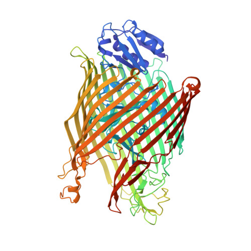
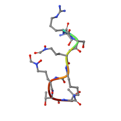
Fpva Bound to Non-Cognate Pyoverdines: Molecular Basis of Siderophore Recognition by an Iron Transporter.

Greenwald, J.Nader, M.Celia, H.Gruffaz, C.Geoffroy, V.Meyer, J.M.Schalk, I.J.Pattus, F.

(2009) Mol Microbiol 72: 1246

  • DOI: https://doi.org/10.1111/j.1365-2958.2009.06721.x
  • Primary Citation of Related Structures:  
    2W16, 2W6T, 2W6U, 2W75, 2W76, 2W77, 2W78

  • PubMed Abstract: 
  • The first step in the specific uptake of iron via siderophores in Gram-negative bacteria is the recognition and binding of a ferric siderophore by its cognate receptor. We investigated the molecular basis of this event through structural and biochemical approaches ...

    The first step in the specific uptake of iron via siderophores in Gram-negative bacteria is the recognition and binding of a ferric siderophore by its cognate receptor. We investigated the molecular basis of this event through structural and biochemical approaches. FpvA, the pyoverdine-Fe transporter from Pseudomonas aeruginosa ATCC 15692 (PAO1 strain), is able to transport ferric-pyoverdines originating from other species, whereas most fluorescent pseudomonads are only able to use the one they produce among the more than 100 known different pyoverdines. We solved the structure of FpvA bound to non-cognate pyoverdines of high- or low-affinity and found a close correlation between receptor-ligand structure and the measured affinities. The structure of the first amino acid residues of the pyoverdine chain distinguished the high- and low-affinity binders while the C-terminal portion of the pyoverdines, often cyclic, does not appear to contribute extensively to the interaction between the siderophore and its transporter. The specificity of the ferric-pyoverdine binding site of FpvA is conferred by the structural elements common to all ferric-pyoverdines, i.e. the chromophore, iron, and its chelating groups.


    Organizational Affiliation

    Laboratoire de Biologie Structurale des Membranes, UMR7175, Ecole Supérieure de Biotechnologie de Strasbourg, Bd Sébastien Brant, BP10413, 67412 Illkirch, France. jason.greenwald@phys.chem.ethz.ch


Macromolecules
Find similar proteins by:  (by identity cutoff)  |  3D Structure
Entity ID: 1
MoleculeChainsSequence LengthOrganismDetailsImage
FERRIPYOVERDINE RECEPTOR
A, B
772Pseudomonas aeruginosa PAO1Mutation(s): 0 
Gene Names: fpvAPA2398
Membrane Entity: Yes 
UniProt
Find proteins for P48632 (Pseudomonas aeruginosa (strain ATCC 15692 / DSM 22644 / CIP 104116 / JCM 14847 / LMG 12228 / 1C / PRS 101 / PAO1))
Explore P48632 
Go to UniProtKB:  P48632
Entity Groups  
Sequence Clusters30% Identity50% Identity70% Identity90% Identity95% Identity100% Identity
UniProt GroupP48632
Protein Feature View
Expand
  • Reference Sequence

Find similar proteins by:  Sequence   |   3D Structure  

Entity ID: 2
MoleculeChainsSequence LengthOrganismDetailsImage
DSN-ARG-DSN-FHO-LYS-FHO-THR-THR8Pseudomonas aeruginosa PAO1Mutation(s): 0 
Protein Feature View
Expand
  • Reference Sequence
Small Molecules
Ligands 4 Unique
IDChainsName / Formula / InChI Key2D Diagram3D Interactions
PVE
Query on PVE

Download Ideal Coordinates CCD File 
P [auth C](1S)-1-CARBOXY-5-[(3-CARBOXYPROPANOYL)AMINO]-8,9-DIHYDROXY-1,2,3,4-TETRAHYDROPYRIMIDO[1,2-A]QUINOLIN-11-IUM
C17 H18 N3 O7
PKIXPGNFUHEBHN-JTQLQIEISA-O
 Ligand Interaction
N8E
Query on N8E

Download Ideal Coordinates CCD File 
D [auth A],
E [auth A]
3,6,9,12,15-PENTAOXATRICOSAN-1-OL
C18 H38 O6
MJELOWOAIAAUJT-UHFFFAOYSA-N
 Ligand Interaction
PO4
Query on PO4

Download Ideal Coordinates CCD File 
F [auth A]
G [auth A]
H [auth A]
I [auth A]
J [auth A]
F [auth A],
G [auth A],
H [auth A],
I [auth A],
J [auth A],
K [auth A],
L [auth B],
M [auth B],
N [auth B],
O [auth B]
PHOSPHATE ION
O4 P
NBIIXXVUZAFLBC-UHFFFAOYSA-K
 Ligand Interaction
FE
Query on FE

Download Ideal Coordinates CCD File 
Q [auth C]FE (III) ION
Fe
VTLYFUHAOXGGBS-UHFFFAOYSA-N
 Ligand Interaction
Modified Residues  1 Unique
IDChainsTypeFormula2D DiagramParent
FHO
Query on FHO
C
L-PEPTIDE LINKINGC6 H12 N2 O4LYS
Biologically Interesting Molecules (External Reference) 1 Unique
Entity ID: 2
IDChainsNameType/Class2D Diagram3D Interactions
PRD_000723
Query on PRD_000723
C
PYOVERDIN C-E Fe ComplexPolypeptide / Metal transport Ligand Interaction
Experimental Data & Validation

Experimental Data

  • Method: X-RAY DIFFRACTION
  • Resolution: 2.71 Å
  • R-Value Free: 0.253 
  • R-Value Work: 0.217 
  • R-Value Observed: 0.219 
  • Space Group: C 1 2 1
Unit Cell:
Length ( Å )Angle ( ˚ )
a = 195.22α = 90
b = 130.082β = 130.67
c = 141.783γ = 90
Software Package:
Software NamePurpose
REFMACrefinement
MOSFLMdata reduction
SCALAdata scaling

Structure Validation

View Full Validation Report


Ligand Structure Quality Assessment 


Entry History 

Deposition Data

Revision History  (Full details and data files)

  • Version 1.0: 2009-05-12
    Type: Initial release
  • Version 1.1: 2011-07-13
    Changes: Advisory, Atomic model, Database references, Derived calculations, Non-polymer description, Refinement description, Structure summary, Version format compliance
  • Version 1.2: 2013-03-06
    Changes: Other
  • Version 1.3: 2018-06-20
    Changes: Data collection, Source and taxonomy
  • Version 1.4: 2019-07-10
    Changes: Data collection, Derived calculations
  • Version 1.5: 2019-07-24
    Changes: Data collection
  • Version 1.6: 2019-07-31
    Changes: Advisory, Data collection, Derived calculations