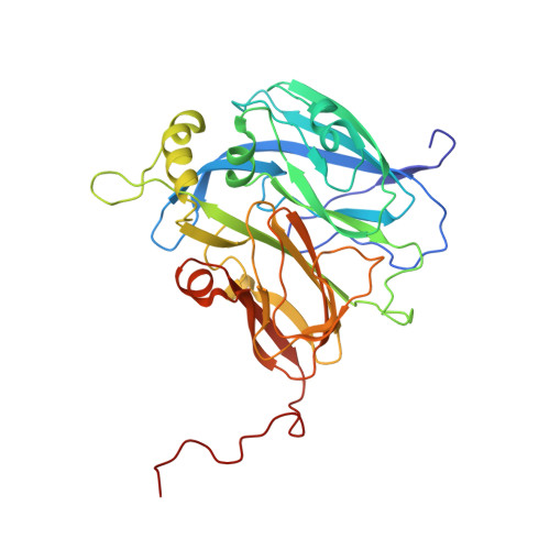
Crystallography with Online Optical and X-Ray Absorption Spectroscopies Demonstrates an Ordered Mechanism in Copper Nitrite Reductase.
Hough, M.A., Antonyuk, S.V., Strange, R.W., Eady, R.R., Hasnain, S.S.(2008) J Mol Biology 378: 353
- PubMed: 18353369
- DOI: https://doi.org/10.1016/j.jmb.2008.01.097
- Primary Citation of Related Structures:
2VM3, 2VM4 - PubMed Abstract:
Nitrite reductases are key enzymes that perform the first committed step in the denitrification process and reduce nitrite to nitric oxide. In copper nitrite reductases, an electron is delivered from the type 1 copper (T1Cu) centre to the type 2 copper (T2Cu) centre where catalysis occurs. Despite significant structural and mechanistic studies, it remains controversial whether the substrates, nitrite, electron and proton are utilised in an ordered or random manner. We have used crystallography, together with online X-ray absorption spectroscopy and optical spectroscopy, to show that X-rays rapidly and selectively photoreduce the T1Cu centre, but that the T2Cu centre does not photoreduce directly over a typical crystallographic data collection time. Furthermore, internal electron transfer between the T1Cu and T2Cu centres does not occur, and the T2Cu centre remains oxidised. These data unambiguously demonstrate an 'ordered' mechanism in which electron transfer is gated by binding of nitrite to the T2Cu. Furthermore, the use of online multiple spectroscopic techniques shows their value in assessing radiation-induced redox changes at different metal sites and demonstrates the importance of ensuring the correct status of redox centres in a crystal structure determination. Here, optical spectroscopy has shown a very high sensitivity for detecting the change in T1Cu redox state, while X-ray absorption spectroscopy has reported on the redox status of the T2Cu site, as this centre has no detectable optical absorption.
- Molecular Biophysics Group, STFC Daresbury Laboratory, Warrington, Cheshire WA4 4AD, UK.
Organizational Affiliation:


