Inhibitors of the Kinase Ispe: Structure-Activity Relationships and Co-Crystal Structure Analysis.
Hirsch, A.K., Alphey, M.S., Lauw, S., Seet, M., Barandun, L., Eisenreich, W., Rohdich, F., Hunter, W.N., Bacher, A., Diederich, F.(2008) Org Biomol Chem 6: 2719
- PubMed: 18633530 Search on PubMed
- DOI: https://doi.org/10.1039/b804375b
- Primary Citation Related Structures:
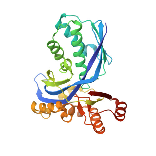
2VF3 - PubMed Abstract:
Enzymes of the non-mevalonate pathway for isoprenoid biosynthesis are therapeutic targets for the treatment of important infectious diseases. Whereas this pathway is absent in humans, it is used by plants, many eubacteria and apicomplexan protozoa, including major human pathogens such as Plasmodium falciparum and Mycobacterium tuberculosis. Herein, we report on the design, preparation and biological evaluation of a new series of ligands for IspE protein, a kinase from this pathway. These inhibitors were developed for the inhibition of IspE from Escherichia coli, using structure-based design approaches. Structure-activity relationships (SARs) and a co-crystal structure of Aquifex aeolicus IspE bound to a representative inhibitor validate the proposed binding mode. The crystal structure shows that the ligand binds in the substrate-rather than the adenosine 5'-triphosphate (ATP)-binding pocket. As predicted, a cyclopropyl substituent occupies a small cavity not used by the substrate. The optimal volume occupancy of this cavity is explored in detail. In the co-crystal structure, a diphosphate anion binds to the Gly-rich loop, which normally accepts the triphosphate moiety of ATP. This structure provides useful insights for future structure-based developments of inhibitors for the parasite enzymes.
- Laboratorium für Organische Chemie, ETH Zürich, HCI, CH-8093, Zürich, Switzerland.
Organizational Affiliation: