2VE7

Crystal structure of a bonsai version of the human Ndc80 complex


Experimental Data Snapshot

  • Method: X-RAY DIFFRACTION
  • Resolution: 2.88 Å
  • R-Value Free: 0.261 
  • R-Value Work: 0.232 
  • R-Value Observed: 0.234 

wwPDB Validation   3D Report Full Report


This is version 1.3 of the entry. See complete history


Literature

Implications for Kinetochore-Microtubule Attachment from the Structure of an Engineered Ndc80 Complex

Ciferri, C.Pasqualato, S.Screpanti, E.Varetti, G.Santaguida, S.Dos Reis, G.Maiolica, A.Polka, J.De Luca, J.G.De Wulf, P.Salek, M.Rappsilber, J.Moores, C.A.Salmon, E.D.Musacchio, A.

(2008) Cell 133: 427

  • DOI: https://doi.org/10.1016/j.cell.2008.03.020
  • Primary Citation of Related Structures:  
    2VE7

  • PubMed Abstract: 

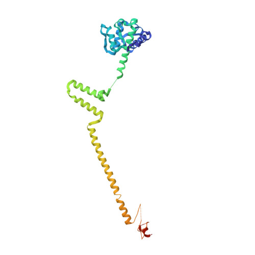
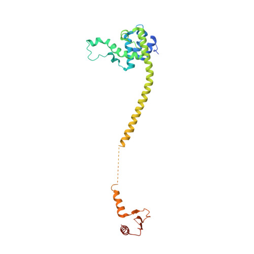
    Kinetochores are proteinaceous assemblies that mediate the interaction of chromosomes with the mitotic spindle. The 180 kDa Ndc80 complex is a direct point of contact between kinetochores and microtubules. Its four subunits contain coiled coils and form an elongated rod structure with functional globular domains at either end. We crystallized an engineered "bonsai" Ndc80 complex containing a shortened rod domain but retaining the globular domains required for kinetochore localization and microtubule binding. The structure reveals a microtubule-binding interface containing a pair of tightly interacting calponin-homology (CH) domains with a previously unknown arrangement. The interaction with microtubules is cooperative and predominantly electrostatic. It involves positive charges in the CH domains and in the N-terminal tail of the Ndc80 subunit and negative charges in tubulin C-terminal tails and is regulated by the Aurora B kinase. We discuss our results with reference to current models of kinetochore-microtubule attachment and centromere organization.


  • Organizational Affiliation

    Department of Experimental Oncology, European Institute of Oncology, Via Adamello 16, I 20139 Milan, Italy.


Macromolecules
Find similar proteins by:  (by identity cutoff)  |  3D Structure
Entity ID: 1
MoleculeChains Sequence LengthOrganismDetailsImage
KINETOCHORE PROTEIN HEC1, KINETOCHORE PROTEIN SPC25
A, B
315Homo sapiensMutation(s): 0 
UniProt & NIH Common Fund Data Resources
Find proteins for Q9HBM1 (Homo sapiens)
Explore Q9HBM1 
Go to UniProtKB:  Q9HBM1
PHAROS:  Q9HBM1
GTEx:  ENSG00000152253 
Find proteins for O14777 (Homo sapiens)
Explore O14777 
Go to UniProtKB:  O14777
PHAROS:  O14777
GTEx:  ENSG00000080986 
Entity Groups  
Sequence Clusters30% Identity50% Identity70% Identity90% Identity95% Identity100% Identity
UniProt GroupsQ9HBM1O14777
Sequence Annotations
Expand
  • Reference Sequence
Find similar proteins by:  (by identity cutoff)  |  3D Structure
Entity ID: 2
MoleculeChains Sequence LengthOrganismDetailsImage
KINETOCHORE PROTEIN NUF2, KINETOCHORE PROTEIN SPC24
C, D
250Homo sapiensMutation(s): 1 
UniProt & NIH Common Fund Data Resources
Find proteins for Q8NBT2 (Homo sapiens)
Explore Q8NBT2 
Go to UniProtKB:  Q8NBT2
PHAROS:  Q8NBT2
GTEx:  ENSG00000161888 
Find proteins for Q9BZD4 (Homo sapiens)
Explore Q9BZD4 
Go to UniProtKB:  Q9BZD4
PHAROS:  Q9BZD4
GTEx:  ENSG00000143228 
Entity Groups  
Sequence Clusters30% Identity50% Identity70% Identity90% Identity95% Identity100% Identity
UniProt GroupsQ8NBT2Q9BZD4
Sequence Annotations
Expand
  • Reference Sequence
Experimental Data & Validation

Experimental Data

  • Method: X-RAY DIFFRACTION
  • Resolution: 2.88 Å
  • R-Value Free: 0.261 
  • R-Value Work: 0.232 
  • R-Value Observed: 0.234 
  • Space Group: P 21 21 2
Unit Cell:
Length ( Å )Angle ( ˚ )
a = 157.653α = 90
b = 248.965β = 90
c = 58.25γ = 90
Software Package:
Software NamePurpose
REFMACrefinement
HKL-2000data reduction
HKL-2000data scaling
SHELXDphasing
SHARPphasing
piratephasing

Structure Validation

View Full Validation Report


Entry History 

Revision History  (Full details and data files)

  • Version 1.0: 2008-05-13
    Type: Initial release
  • Version 1.1: 2011-07-13
    Changes: Refinement description, Version format compliance
  • Version 1.2: 2017-06-21
    Changes: Refinement description
  • Version 1.3: 2022-05-04
    Changes: Advisory, Database references