2V5P

COMPLEX STRUCTURE OF HUMAN IGF2R DOMAINS 11-13 BOUND TO IGF-II


Experimental Data Snapshot

  • Method: X-RAY DIFFRACTION
  • Resolution: 4.10 Å
  • R-Value Free: 0.327 
  • R-Value Work: 0.291 
  • R-Value Observed: 0.293 

wwPDB Validation   3D Report Full Report


Ligand Structure Quality Assessment 


This is version 2.0 of the entry. See complete history


Literature

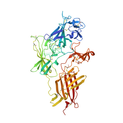
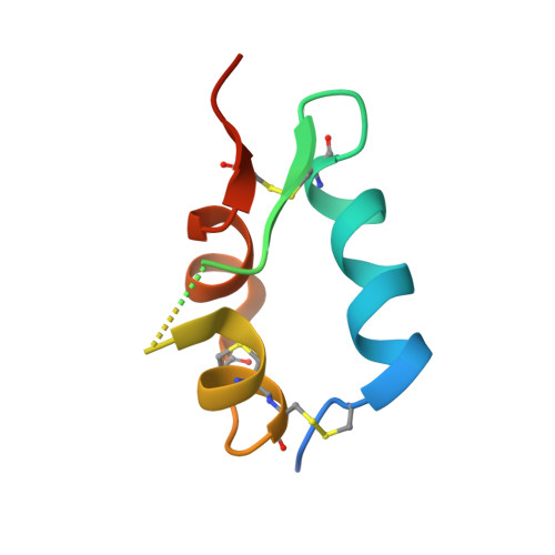
Structure and Functional Analysis of the Igf-II/Igf2R Interaction

Brown, J.Delaine, C.Zaccheo, O.J.Siebold, C.Gilbert, R.J.Van Boxel, G.Denley, A.Wallace, J.C.Hassan, A.B.Forbes, B.E.Jones, E.Y.

(2008) EMBO J 27: 265

  • DOI: https://doi.org/10.1038/sj.emboj.7601938
  • Primary Citation of Related Structures:  
    2V5N, 2V5O, 2V5P

  • PubMed Abstract: 
  • Embryonic development and normal growth require exquisite control of insulin-like growth factors (IGFs). In mammals the extracellular region of the cation-independent mannose-6-phosphate receptor has gained an IGF-II-binding function and is termed type II IGF receptor (IGF2R) ...

    Embryonic development and normal growth require exquisite control of insulin-like growth factors (IGFs). In mammals the extracellular region of the cation-independent mannose-6-phosphate receptor has gained an IGF-II-binding function and is termed type II IGF receptor (IGF2R). IGF2R sequesters IGF-II; imbalances occur in cancers and IGF2R is implicated in tumour suppression. We report crystal structures of IGF2R domains 11-12, 11-12-13-14 and domains 11-12-13/IGF-II complex. A distinctive juxtaposition of these domains provides the IGF-II-binding unit, with domain 11 directly interacting with IGF-II and domain 13 modulating binding site flexibility. Our complex shows that Phe19 and Leu53 of IGF-II lock into a hydrophobic pocket unique to domain 11 of mammalian IGF2Rs. Mutagenesis analyses confirm this IGF-II 'binding-hotspot', revealing that IGF-binding proteins and IGF2R have converged on the same high-affinity site.


    Organizational Affiliation

    Cancer Research UK Receptor Structure Research Group, Division of Structural Biology, Wellcome Trust Centre for Human Genetics, University of Oxford, Headington, Oxford, UK.


Macromolecules
Find similar proteins by:  (by identity cutoff)  |  3D Structure
Entity ID: 1
MoleculeChainsSequence LengthOrganismDetailsImage
CATION-INDEPENDENT MANNOSE-6-PHOSPHATE RECEPTORA, B492Homo sapiensMutation(s): 0 
Gene Names: IGF2RMPRI
UniProt & NIH Common Fund Data Resources
Find proteins for P11717 (Homo sapiens)
Explore P11717 
Go to UniProtKB:  P11717
PHAROS:  P11717
Entity Groups  
Sequence Clusters30% Identity50% Identity70% Identity90% Identity95% Identity100% Identity
UniProt GroupP11717
Protein Feature View
Expand
  • Reference Sequence
Find similar proteins by:  (by identity cutoff)  |  3D Structure
Entity ID: 2
MoleculeChainsSequence LengthOrganismDetailsImage
INSULIN-LIKE GROWTH FACTOR IIC, D67Homo sapiensMutation(s): 0 
Gene Names: IGF2PP1446
UniProt & NIH Common Fund Data Resources
Find proteins for P01344 (Homo sapiens)
Explore P01344 
Go to UniProtKB:  P01344
PHAROS:  P01344
Entity Groups  
Sequence Clusters30% Identity50% Identity70% Identity90% Identity95% Identity100% Identity
UniProt GroupP01344
Protein Feature View
Expand
  • Reference Sequence
Oligosaccharides

Help

Entity ID: 3
MoleculeChainsChain Length2D DiagramGlycosylation3D Interactions
beta-D-mannopyranose-(1-4)-2-acetamido-2-deoxy-beta-D-glucopyranose-(1-4)-2-acetamido-2-deoxy-beta-D-glucopyranoseE 3N-Glycosylation Oligosaccharides Interaction
Glycosylation Resources
GlyTouCan:  G15407YE
GlyCosmos:  G15407YE
GlyGen:  G15407YE
Small Molecules
Ligands 1 Unique
IDChainsName / Formula / InChI Key2D Diagram3D Interactions
NAG
Query on NAG

Download Ideal Coordinates CCD File 
F [auth A],
G [auth A],
H [auth B],
I [auth B],
J [auth B]
2-acetamido-2-deoxy-beta-D-glucopyranose
C8 H15 N O6
OVRNDRQMDRJTHS-FMDGEEDCSA-N
 Ligand Interaction
Experimental Data & Validation

Experimental Data

  • Method: X-RAY DIFFRACTION
  • Resolution: 4.10 Å
  • R-Value Free: 0.327 
  • R-Value Work: 0.291 
  • R-Value Observed: 0.293 
  • Space Group: C 1 2 1
Unit Cell:
Length ( Å )Angle ( ˚ )
a = 165.979α = 90
b = 117.312β = 123.41
c = 116.668γ = 90
Software Package:
Software NamePurpose
REFMACrefinement
XDSdata reduction
XDSdata scaling
MOLREPphasing

Structure Validation

View Full Validation Report


Ligand Structure Quality Assessment 


Entry History 

Deposition Data

Revision History  (Full details and data files)

  • Version 1.0: 2007-12-11
    Type: Initial release
  • Version 1.1: 2011-05-08
    Changes: Version format compliance
  • Version 1.2: 2011-07-13
    Changes: Version format compliance
  • Version 1.3: 2018-02-28
    Changes: Source and taxonomy
  • Version 2.0: 2020-07-29
    Type: Remediation
    Reason: Carbohydrate remediation
    Changes: Atomic model, Data collection, Derived calculations, Other, Structure summary