Crystallographic and Spectroscopic Studies of Peroxide-Derived Myoglobin Compound II and Occurrence of Protonated Fe(Iv)-O
Hersleth, H.-P., Uchida, T., Rohr, A.K., Teschner, T., Schunemann, V., Kitagawa, T., Trautwein, A.X., Gorbitz, C.H., Andersson, K.K.(2007) J Biological Chem 282: 23372
- PubMed: 17565988 Search on PubMed
- DOI: https://doi.org/10.1074/jbc.M701948200
- Primary Citation Related Structures:
2V1E, 2V1F, 2V1G, 2V1H, 2V1I, 2V1J, 2V1K - PubMed Abstract:
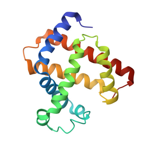
High resolution crystal structures of myoglobin in the pH range 5.2-8.7 have been used as models for the peroxide-derived compound II intermediates in heme peroxidases and oxygenases. The observed Fe-O bond length (1.86-1.90 A) is consistent with that of a single bond. The compound II state of myoglobin in crystals was controlled by single-crystal microspectrophotometry before and after synchrotron data collection. We observe some radiation-induced changes in both compound II (resulting in intermediate H) and in the resting ferric state of myoglobin. These radiation-induced states are quite unstable, and compound II and ferric myoglobin are immediately regenerated through a short heating above the glass transition temperature (<1 s) of the crystals. It is unclear how this influences our compound II structures compared with the unaffected compound II, but some crystallographic data suggest that the influence on the Fe-O bond distance is minimal. Based on our crystallographic and spectroscopic data we suggest that for myoglobin the compound II intermediate consists of an Fe(IV)-O species with a single bond. The presence of Fe(IV) is indicated by a small isomer shift of delta = 0.07 mm/s from Mössbauer spectroscopy. Earlier quantum refinements (crystallographic refinement where the molecular-mechanics potential is replaced by a quantum chemical calculation) and density functional theory calculations suggest that this intermediate H species is protonated.
- Department of Chemistry, University of Oslo, PO Box 1033, Blindern, Oslo N-0315, Norway.
Organizational Affiliation: