2THF

STRUCTURE OF HUMAN ALPHA-THROMBIN Y225F MUTANT BOUND TO D-PHE-PRO-ARG-CHLOROMETHYLKETONE


Experimental Data Snapshot

  • Method: X-RAY DIFFRACTION
  • Resolution: 2.10 Å
  • R-Value Free: 0.265 
  • R-Value Work: 0.199 

wwPDB Validation   3D Report Full Report


This is version 1.4 of the entry. See complete history


Literature

Unexpected crucial role of residue 225 in serine proteases.

Guinto, E.R.Caccia, S.Rose, T.Futterer, K.Waksman, G.Di Cera, E.

(1999) Proc Natl Acad Sci U S A 96: 1852-1857

  • DOI: https://doi.org/10.1073/pnas.96.5.1852
  • Primary Citation of Related Structures:  
    1B7X, 1THP, 2THF

  • PubMed Abstract: 

    Residue 225 in serine proteases of the chymotrypsin family is Pro or Tyr in more than 95% of nearly 300 available sequences. Proteases with Y225 (like some blood coagulation and complement factors) are almost exclusively found in vertebrates, whereas proteases with P225 (like degradative enzymes) are present from bacteria to human. Saturation mutagenesis of Y225 in thrombin shows that residue 225 affects ligand recognition up to 60,000-fold. With the exception of Tyr and Phe, all residues are associated with comparable or greatly reduced catalytic activity relative to Pro. The crystal structures of three mutants that differ widely in catalytic activity (Y225F, Y225P, and Y225I) show that although residue 225 makes no contact with substrate, it drastically influences the shape of the water channel around the primary specificity site. The activity profiles obtained for thrombin also suggest that the conversion of Pro to Tyr or Phe documented in the vertebrates occurred through Ser and was driven by a significant gain (up to 50-fold) in catalytic activity. In fact, Ser and Phe are documented in 4% of serine proteases, which together with Pro and Tyr account for almost the entire distribution of residues at position 225. The unexpected crucial role of residue 225 in serine proteases explains the evolutionary selection of residues at this position and shows that the structural determinants of protease activity and specificity are more complex than currently believed. These findings have broad implications in the rational design of enzymes with enhanced catalytic properties.


  • Organizational Affiliation

    Department of Biochemistry and Molecular Biophysics, Washington University School of Medicine, Box 8231, St. Louis, MO 63110, USA.


Macromolecules
Find similar proteins by:  (by identity cutoff)  |  3D Structure
Entity ID: 1
MoleculeChains Sequence LengthOrganismDetailsImage
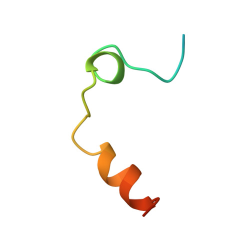
THROMBIN LIGHT CHAIN36Homo sapiensMutation(s): 0 
UniProt & NIH Common Fund Data Resources
Find proteins for P00734 (Homo sapiens)
Explore P00734 
Go to UniProtKB:  P00734
PHAROS:  P00734
GTEx:  ENSG00000180210 
Entity Groups  
Sequence Clusters30% Identity50% Identity70% Identity90% Identity95% Identity100% Identity
UniProt GroupP00734
Sequence Annotations
Expand
  • Reference Sequence
Find similar proteins by:  (by identity cutoff)  |  3D Structure
Entity ID: 2
MoleculeChains Sequence LengthOrganismDetailsImage
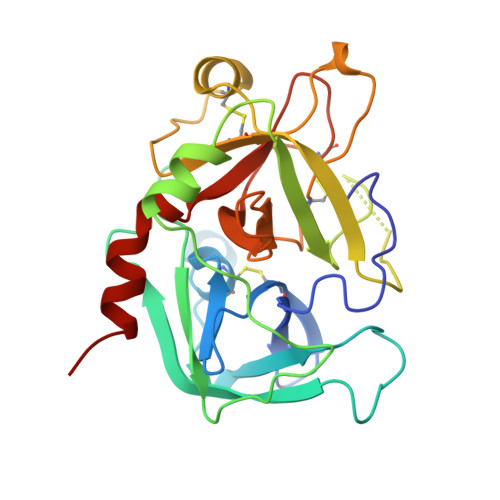
THROMBIN HEAVY CHAIN259Homo sapiensMutation(s): 0 
EC: 3.4.21.5
UniProt & NIH Common Fund Data Resources
Find proteins for P00734 (Homo sapiens)
Explore P00734 
Go to UniProtKB:  P00734
PHAROS:  P00734
GTEx:  ENSG00000180210 
Entity Groups  
Sequence Clusters30% Identity50% Identity70% Identity90% Identity95% Identity100% Identity
UniProt GroupP00734
Sequence Annotations
Expand
  • Reference Sequence
Small Molecules
Ligands 2 Unique
IDChains Name / Formula / InChI Key2D Diagram3D Interactions
0G6
Query on 0G6

Download Ideal Coordinates CCD File 
C [auth B]D-phenylalanyl-N-[(2S,3S)-6-{[amino(iminio)methyl]amino}-1-chloro-2-hydroxyhexan-3-yl]-L-prolinamide
C21 H34 Cl N6 O3
DVFLYEYCMMLBTQ-VSZNYVQBSA-O
NA
Query on NA

Download Ideal Coordinates CCD File 
D [auth B]SODIUM ION
Na
FKNQFGJONOIPTF-UHFFFAOYSA-N
Biologically Interesting Molecules (External Reference) 1 Unique
Entity ID: 3
IDChains NameType/Class2D Diagram3D Interactions
PRD_000020 (0G6)
Query on PRD_000020
C [auth B]D-Phe-Pro-Arg-CH2ClPeptide-like / Inhibitor
Experimental Data & Validation

Experimental Data

  • Method: X-RAY DIFFRACTION
  • Resolution: 2.10 Å
  • R-Value Free: 0.265 
  • R-Value Work: 0.199 
  • Space Group: P 21 21 21
Unit Cell:
Length ( Å )Angle ( ˚ )
a = 53.1α = 90
b = 75.1β = 90
c = 81.4γ = 90
Software Package:
Software NamePurpose
DENZOdata reduction
SCALEPACKdata scaling
AMoREphasing
X-PLORrefinement

Structure Validation

View Full Validation Report


Entry History 

Deposition Data

Revision History  (Full details and data files)

  • Version 1.0: 1999-03-07
    Type: Initial release
  • Version 1.1: 2008-04-26
    Changes: Version format compliance
  • Version 1.2: 2011-07-13
    Changes: Atomic model, Database references, Derived calculations, Non-polymer description, Structure summary, Version format compliance
  • Version 1.3: 2013-02-27
    Changes: Other
  • Version 1.4: 2023-08-30
    Changes: Data collection, Database references, Derived calculations, Refinement description