2SBL

THE THREE-DIMENSIONAL STRUCTURE OF AN ARACHIDONIC ACID 15-LIPOXYGENASE


Experimental Data Snapshot

  • Method: X-RAY DIFFRACTION
  • Resolution: 2.60 Å
  • R-Value Work: 0.173 
  • R-Value Observed: 0.173 

wwPDB Validation   3D Report Full Report


This is version 1.3 of the entry. See complete history


Literature

The three-dimensional structure of an arachidonic acid 15-lipoxygenase.

Boyington, J.C.Gaffney, B.J.Amzel, L.M.

(1993) Science 260: 1482-1486

  • DOI: https://doi.org/10.1126/science.8502991
  • Primary Citation of Related Structures:  
    2SBL

  • PubMed Abstract: 

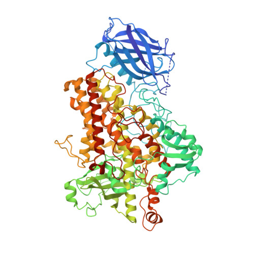
    In mammals, the hydroperoxidation of arachidonic acid by lipoxygenases leads to the formation of leukotrienes and lipoxins, compounds that mediate inflammatory responses. Lipoxygenases are dioxygenases that contain a nonheme iron and are present in many animal cells. Soybean lipoxygenase-1 is a single-chain, 839-residue protein closely related to mammalian lipoxygenases. The structure of soybean lipoxygenase-1 solved to 2.6 angstrom resolution shows that the enzyme has two domains: a 146-residue beta barrel and a 693-residue helical bundle. The iron atom is in the center of the larger domain and is coordinated by three histidines and the COO- of the carboxyl terminus. The coordination geometry is nonregular and appears to be a distorted octahedron in which two adjacent positions are not occupied by ligands. Two cavities, in the shapes of a bent cylinder and a frustum, connect the unoccupied positions to the surface of the enzyme. The iron, with two adjacent and unoccupied positions, is poised to interact with the 1,4-diene system of the substrate and with molecular oxygen during catalysis.


  • Organizational Affiliation

    Department of Biophysics and Biophysical Chemistry, Johns Hopkins University School of Medicine, Baltimore, MD 21205.


Macromolecules
Find similar proteins by:  (by identity cutoff)  |  3D Structure
Entity ID: 1
MoleculeChains Sequence LengthOrganismDetailsImage
LIPOXYGENASE-1A [auth B],
B [auth A]
839Glycine maxMutation(s): 0 
EC: 1.13.11.12
UniProt
Find proteins for P08170 (Glycine max)
Explore P08170 
Go to UniProtKB:  P08170
Entity Groups  
Sequence Clusters30% Identity50% Identity70% Identity90% Identity95% Identity100% Identity
UniProt GroupP08170
Sequence Annotations
Expand
  • Reference Sequence
Small Molecules
Ligands 1 Unique
IDChains Name / Formula / InChI Key2D Diagram3D Interactions
FE
Query on FE

Download Ideal Coordinates CCD File 
C [auth B],
D [auth A]
FE (III) ION
Fe
VTLYFUHAOXGGBS-UHFFFAOYSA-N
Experimental Data & Validation

Experimental Data

  • Method: X-RAY DIFFRACTION
  • Resolution: 2.60 Å
  • R-Value Work: 0.173 
  • R-Value Observed: 0.173 
  • Space Group: C 1 2 1
Unit Cell:
Length ( Å )Angle ( ˚ )
a = 184.5α = 90
b = 125.6β = 102.9
c = 94.7γ = 90
Software Package:
Software NamePurpose
X-PLORmodel building
X-PLORrefinement
X-PLORphasing

Structure Validation

View Full Validation Report


Entry History 

Deposition Data

Revision History  (Full details and data files)

  • Version 1.0: 1995-02-27
    Type: Initial release
  • Version 1.1: 2008-03-25
    Changes: Version format compliance
  • Version 1.2: 2011-07-13
    Changes: Version format compliance
  • Version 1.3: 2024-02-21
    Changes: Data collection, Database references, Derived calculations, Other