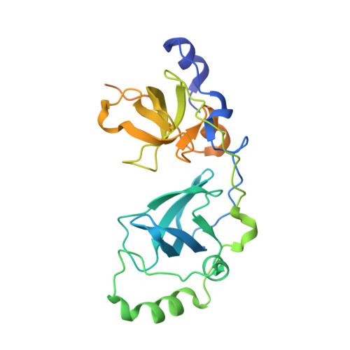
2R58

Crystal Structure of the two MBT repeats from Sex-Comb on Midleg (SCM) in Complex with Di-Methyl Lysine


Experimental Data Snapshot

  • Method: X-RAY DIFFRACTION
  • Resolution: 2.00 Å
  • R-Value Free: 0.229 
  • R-Value Work: 0.186 
  • R-Value Observed: 0.188 

Starting Model: experimental
View more details

wwPDB Validation   3D Report Full Report


Ligand Structure Quality Assessment 


This is version 1.3 of the entry. See complete history


Literature

Structural and functional analyses of methyl-lysine binding by the malignant brain tumour repeat protein Sex comb on midleg.

Grimm, C.de Ayala Alonso, A.G.Rybin, V.Steuerwald, U.Ly-Hartig, N.Fischle, W.Muller, J.Muller, C.W.

(2007) EMBO Rep 8: 1031-1037

  • DOI: https://doi.org/10.1038/sj.embor.7401085
  • Primary Citation of Related Structures:  
    2R57, 2R58, 2R5A, 2R5M

  • PubMed Abstract: 

    Sex comb on midleg (Scm) is a member of the Polycomb group of proteins involved in the maintenance of repression of Hox and other developmental control genes in Drosophila. The two malignant brain tumour (MBT) repeats of Scm form a domain that preferentially binds to monomethylated lysine residues either as a free amino acid or in the context of peptides, while unmodified or di- or trimethylated lysine residues are bound with significantly lower affinity. The crystal structure of a monomethyl-lysine-containing histone tail peptide bound to the MBT repeat domain shows that the methyl-lysine side chain occupies a binding pocket in the second MBT repeat formed by three conserved aromatic residues and one aspartate. Insertion of the monomethylated side chain into this pocket seems to be the main contributor to the binding affinity. Functional analyses in Drosophila show that the MBT domain of Scm and its methyl-lysine-binding activity are required for repression of Hox genes.


  • Organizational Affiliation

    European Molecular Biology Laboratory, Grenoble Outstation, 6 Rue Jules Horowitz, 38042 Grenoble, France.


Macromolecules
Find similar proteins by:  (by identity cutoff)  |  3D Structure
Entity ID: 1
MoleculeChains Sequence LengthOrganismDetailsImage
Polycomb protein Scm265Drosophila melanogasterMutation(s): 1 
Gene Names: Scm
UniProt
Find proteins for Q9VHA0 (Drosophila melanogaster)
Explore Q9VHA0 
Go to UniProtKB:  Q9VHA0
Entity Groups  
Sequence Clusters30% Identity50% Identity70% Identity90% Identity95% Identity100% Identity
UniProt GroupQ9VHA0
Sequence Annotations
Expand
  • Reference Sequence
Small Molecules
Ligands 1 Unique
IDChains Name / Formula / InChI Key2D Diagram3D Interactions
MLY
Query on MLY

Download Ideal Coordinates CCD File 
B [auth A]N-DIMETHYL-LYSINE
C8 H18 N2 O2
XXEWFEBMSGLYBY-ZETCQYMHSA-N
Binding Affinity Annotations 
IDSourceBinding Affinity
MLY PDBBind:  2R58 Kd: 1.00e+7 (nM) from 1 assay(s)
Experimental Data & Validation

Experimental Data

  • Method: X-RAY DIFFRACTION
  • Resolution: 2.00 Å
  • R-Value Free: 0.229 
  • R-Value Work: 0.186 
  • R-Value Observed: 0.188 
  • Space Group: P 43 21 2
Unit Cell:
Length ( Å )Angle ( ˚ )
a = 83.12α = 90
b = 83.12β = 90
c = 75.768γ = 90
Software Package:
Software NamePurpose
REFMACrefinement
DNAdata collection
XDSdata reduction
XSCALEdata scaling
PHASERphasing

Structure Validation

View Full Validation Report


Ligand Structure Quality Assessment 


Entry History 

Deposition Data

Revision History  (Full details and data files)

  • Version 1.0: 2007-10-30
    Type: Initial release
  • Version 1.1: 2011-07-13
    Changes: Advisory, Version format compliance
  • Version 1.2: 2021-10-20
    Changes: Database references, Derived calculations
  • Version 1.3: 2023-08-30
    Changes: Data collection, Refinement description