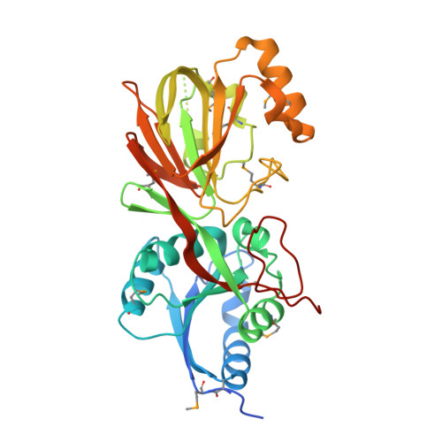
2QV7

Crystal Structure of Diacylglycerol Kinase DgkB in complex with ADP and Mg


Experimental Data Snapshot

  • Method: X-RAY DIFFRACTION
  • Resolution: 2.30 Å
  • R-Value Free: 0.246 
  • R-Value Work: 0.206 
  • R-Value Observed: 0.208 

wwPDB Validation   3D Report Full Report


Ligand Structure Quality Assessment 


This is version 1.1 of the entry. See complete history


Literature

Analysis of the Staphylococcus aureus DgkB Structure Reveals a Common Catalytic Mechanism for the Soluble Diacylglycerol Kinases.

Miller, D.J.Jerga, A.Rock, C.O.White, S.W.

(2008) Structure 16: 1036-1046

  • DOI: https://doi.org/10.1016/j.str.2008.03.019
  • Primary Citation of Related Structures:  
    2QV7, 2QVL

  • PubMed Abstract: 

    Soluble diacylglycerol (DAG) kinases function as regulators of diacylglycerol metabolism in cell signaling and intermediary metabolism. We report the structure of a DAG kinase, DgkB from Staphylococcus aureus, both as the free enzyme and in complex with ADP. The molecule is a tight homodimer, and each monomer comprises two domains with the catalytic center located within the interdomain cleft. Two distinctive features of DkgB are a structural Mg2+ site and an associated Asp*water*Mg2+ network that extends toward the active site locale. Site-directed mutagenesis revealed that these features play important roles in the catalytic mechanism. The key active site residues and the components of the Asp*water*Mg2+ network are conserved in the catalytic cores of the mammalian signaling DAG kinases, indicating that these enzymes use the same mechanism and have similar structures as DgkB.


  • Organizational Affiliation

    Department of Structural Biology, St. Jude Children's Research Hospital, and Department of Molecular Sciences, University of Tennessee Health Science Center, 332 North Lauderdale Street, Memphis, TN 38105, USA.


Macromolecules
Find similar proteins by:  (by identity cutoff)  |  3D Structure
Entity ID: 1
MoleculeChains Sequence LengthOrganismDetailsImage
Diacylglycerol Kinase DgkB337Staphylococcus aureusMutation(s): 0 
Gene Names: SAR1989
EC: 2.7.1.107
UniProt
Find proteins for Q6GFF9 (Staphylococcus aureus (strain MRSA252))
Explore Q6GFF9 
Go to UniProtKB:  Q6GFF9
Entity Groups  
Sequence Clusters30% Identity50% Identity70% Identity90% Identity95% Identity100% Identity
UniProt GroupQ6GFF9
Sequence Annotations
Expand
  • Reference Sequence
Small Molecules
Ligands 2 Unique
IDChains Name / Formula / InChI Key2D Diagram3D Interactions
ADP
Query on ADP

Download Ideal Coordinates CCD File 
C [auth A]ADENOSINE-5'-DIPHOSPHATE
C10 H15 N5 O10 P2
XTWYTFMLZFPYCI-KQYNXXCUSA-N
MG
Query on MG

Download Ideal Coordinates CCD File 
B [auth A]MAGNESIUM ION
Mg
JLVVSXFLKOJNIY-UHFFFAOYSA-N
Modified Residues  1 Unique
IDChains TypeFormula2D DiagramParent
MSE
Query on MSE
A
L-PEPTIDE LINKINGC5 H11 N O2 SeMET
Binding Affinity Annotations 
IDSourceBinding Affinity
ADP PDBBind:  2QV7 Kd: 2.19e+5 (nM) from 1 assay(s)
Experimental Data & Validation

Experimental Data

  • Method: X-RAY DIFFRACTION
  • Resolution: 2.30 Å
  • R-Value Free: 0.246 
  • R-Value Work: 0.206 
  • R-Value Observed: 0.208 
  • Space Group: P 42 21 2
Unit Cell:
Length ( Å )Angle ( ˚ )
a = 123.76α = 90
b = 123.76β = 90
c = 47.68γ = 90
Software Package:
Software NamePurpose
REFMACrefinement
HKL-2000data collection
HKL-2000data reduction
HKL-2000data scaling
DgkBphasing

Structure Validation

View Full Validation Report


Ligand Structure Quality Assessment 


Entry History 

Deposition Data

Revision History  (Full details and data files)

  • Version 1.0: 2008-06-17
    Type: Initial release
  • Version 1.1: 2011-07-13
    Changes: Advisory, Refinement description, Version format compliance