2QT4

Atomic-resolution crystal structure of the natural form of Scytovirin


Experimental Data Snapshot

  • Method: X-RAY DIFFRACTION
  • Resolution: 1.30 Å
  • R-Value Free: 0.198 
  • R-Value Work: 0.145 
  • R-Value Observed: 0.147 

wwPDB Validation   3D Report Full Report


This is version 1.2 of the entry. See complete history


Literature

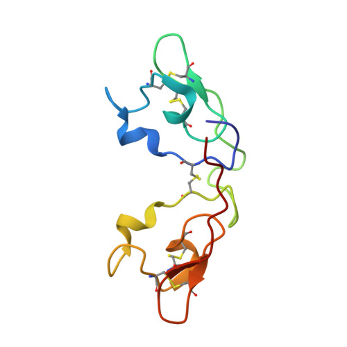
Atomic-resolution crystal structure of the antiviral lectin scytovirin.

Moulaei, T.Botos, I.Ziolkowska, N.E.Bokesch, H.R.Krumpe, L.R.McKee, T.C.O'Keefe, B.R.Dauter, Z.Wlodawer, A.

(2007) Protein Sci 16: 2756-2760

  • DOI: https://doi.org/10.1110/ps.073157507
  • Primary Citation of Related Structures:  
    2QSK, 2QT4

  • PubMed Abstract: 

    The crystal structures of the natural and recombinant antiviral lectin scytovirin (SVN) were solved by single-wavelength anomalous scattering and refined with data extending to 1.3 A and 1.0 A resolution, respectively. A molecule of SVN consists of a single chain 95 amino acids long, with an almost perfect sequence repeat that creates two very similar domains (RMS deviation 0.25 A for 40 pairs of Calpha atoms). The crystal structure differs significantly from a previously published NMR structure of the same protein, with the RMS deviations calculated separately for the N- and C-terminal domains of 5.3 A and 3.7 A, respectively, and a very different relationship between the two domains. In addition, the disulfide bonding pattern of the crystal structures differs from that described in the previously published mass spectrometry and NMR studies.


  • Organizational Affiliation

    Protein Structure Section, Macromolecular Crystallography Laboratory, National Cancer Institute, NCI-Frederick, Frederick, Maryland 21702-1201, USA.


Macromolecules
Find similar proteins by:  (by identity cutoff)  |  3D Structure
Entity ID: 1
MoleculeChains Sequence LengthOrganismDetailsImage
scytovirin95Scytonema variumMutation(s): 0 
UniProt
Find proteins for P86041 (Scytonema varium)
Explore P86041 
Go to UniProtKB:  P86041
Entity Groups  
Sequence Clusters30% Identity50% Identity70% Identity90% Identity95% Identity100% Identity
UniProt GroupP86041
Sequence Annotations
Expand
  • Reference Sequence
Experimental Data & Validation

Experimental Data

  • Method: X-RAY DIFFRACTION
  • Resolution: 1.30 Å
  • R-Value Free: 0.198 
  • R-Value Work: 0.145 
  • R-Value Observed: 0.147 
  • Space Group: P 21 21 21
Unit Cell:
Length ( Å )Angle ( ˚ )
a = 37.48α = 90
b = 38.84β = 90
c = 59.72γ = 90
Software Package:
Software NamePurpose
DENZOdata reduction
SCALEPACKdata scaling
REFMACrefinement
PDB_EXTRACTdata extraction
HKL-2000data collection
SHELXSphasing

Structure Validation

View Full Validation Report


Entry History 

Deposition Data

Revision History  (Full details and data files)

  • Version 1.0: 2007-11-27
    Type: Initial release
  • Version 1.1: 2011-07-13
    Changes: Version format compliance
  • Version 1.2: 2017-10-25
    Changes: Refinement description