2Q6U

SeMet-substituted form of NikD


Experimental Data Snapshot

  • Method: X-RAY DIFFRACTION
  • Resolution: 1.75 Å
  • R-Value Free: 0.228 
  • R-Value Work: 0.191 

wwPDB Validation   3D Report Full Report


Ligand Structure Quality Assessment 


This is version 1.2 of the entry. See complete history


Literature

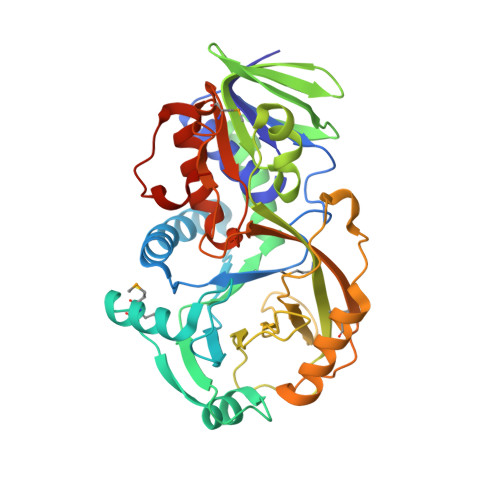
NikD, an Unusual Amino Acid Oxidase Essential for Nikkomycin Biosynthesis: Structures of Closed and Open Forms at 1.15 and 1.90 A Resolution

Carrell, C.J.Bruckner, R.C.Venci, D.Zhao, G.Jorns, M.S.Mathews, F.S.

(2007) Structure 15: 928-941

  • DOI: https://doi.org/10.1016/j.str.2007.06.010
  • Primary Citation of Related Structures:  
    2OLN, 2OLO, 2Q6U

  • PubMed Abstract: 

    NikD is an unusual amino-acid-oxidizing enzyme that contains covalently bound FAD, catalyzes a 4-electron oxidation of piperideine-2-carboxylic acid to picolinate, and plays a critical role in the biosynthesis of nikkomycin antibiotics. Crystal structures of closed and open forms of nikD, a two-domain enzyme, have been determined to resolutions of 1.15 and 1.9 A, respectively. The two forms differ by an 11 degrees rotation of the catalytic domain with respect to the FAD-binding domain. The active site is inaccessible to solvent in the closed form; an endogenous ligand, believed to be picolinate, is bound close to and parallel with the flavin ring, an orientation compatible with redox catalysis. The active site is solvent accessible in the open form, but the picolinate ligand is approximately perpendicular to the flavin ring and a tryptophan is stacked above the flavin ring. NikD also contains a mobile cation binding loop.


  • Organizational Affiliation

    Department of Biochemistry and Molecular Biophysics, Washington University School of Medicine, St. Louis, MO 63110, USA.


Macromolecules
Find similar proteins by:  (by identity cutoff)  |  3D Structure
Entity ID: 1
MoleculeChains Sequence LengthOrganismDetailsImage
NikD protein395Streptomyces tendaeMutation(s): 1 
Gene Names: nikD
UniProt
Find proteins for Q9X9P9 (Streptomyces tendae)
Explore Q9X9P9 
Go to UniProtKB:  Q9X9P9
Entity Groups  
Sequence Clusters30% Identity50% Identity70% Identity90% Identity95% Identity100% Identity
UniProt GroupQ9X9P9
Sequence Annotations
Expand
  • Reference Sequence
Small Molecules
Ligands 2 Unique
IDChains Name / Formula / InChI Key2D Diagram3D Interactions
FAD
Query on FAD

Download Ideal Coordinates CCD File 
B [auth A]FLAVIN-ADENINE DINUCLEOTIDE
C27 H33 N9 O15 P2
VWWQXMAJTJZDQX-UYBVJOGSSA-N
BEZ
Query on BEZ

Download Ideal Coordinates CCD File 
C [auth A]BENZOIC ACID
C7 H6 O2
WPYMKLBDIGXBTP-UHFFFAOYSA-N
Modified Residues  1 Unique
IDChains TypeFormula2D DiagramParent
MSE
Query on MSE
A
L-PEPTIDE LINKINGC5 H11 N O2 SeMET
Experimental Data & Validation

Experimental Data

  • Method: X-RAY DIFFRACTION
  • Resolution: 1.75 Å
  • R-Value Free: 0.228 
  • R-Value Work: 0.191 
  • Space Group: C 1 2 1
Unit Cell:
Length ( Å )Angle ( ˚ )
a = 78.52α = 90
b = 96.12β = 118.49
c = 78.05γ = 90
Software Package:
Software NamePurpose
DENZOdata reduction
SCALEPACKdata scaling
CNSrefinement
PDB_EXTRACTdata extraction
MAR345dtbdata collection
HKL-2000data reduction
XFITdata reduction

Structure Validation

View Full Validation Report


Ligand Structure Quality Assessment 


Entry History 

Deposition Data

Revision History  (Full details and data files)

  • Version 1.0: 2007-07-31
    Type: Initial release
  • Version 1.1: 2011-07-13
    Changes: Version format compliance
  • Version 1.2: 2017-10-18
    Changes: Refinement description