2Q4G

Ensemble refinement of the protein crystal structure of human ribonuclease inhibitor complexed with ribonuclease I


Experimental Data Snapshot

  • Method: X-RAY DIFFRACTION
  • Resolution: 1.95 Å
  • R-Value Free: 0.224 
  • R-Value Work: 0.161 
  • R-Value Observed: 0.161 

Starting Model: experimental
View more details

wwPDB Validation   3D Report Full Report


This is version 1.4 of the entry. See complete history

Re-refinement Note

This entry reflects an alternative modeling of the original data in: 1Z7X


Literature

Inhibition of human pancreatic ribonuclease by the human ribonuclease inhibitor protein.

Johnson, R.J.McCoy, J.G.Bingman, C.A.Phillips Jr., G.N.Raines, R.T.

(2007) J Mol Biol 368: 434-449

  • DOI: https://doi.org/10.1016/j.jmb.2007.02.005
  • Primary Citation of Related Structures:  
    1Z7X, 2Q4G

  • PubMed Abstract: 

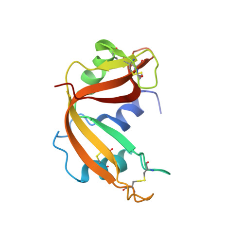
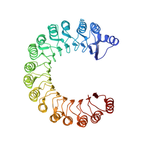
    The ribonuclease inhibitor protein (RI) binds to members of the bovine pancreatic ribonuclease (RNase A) superfamily with an affinity in the femtomolar range. Here, we report on structural and energetic aspects of the interaction between human RI (hRI) and human pancreatic ribonuclease (RNase 1). The structure of the crystalline hRI x RNase 1 complex was determined at a resolution of 1.95 A, revealing the formation of 19 intermolecular hydrogen bonds involving 13 residues of RNase 1. In contrast, only nine such hydrogen bonds are apparent in the structure of the complex between porcine RI and RNase A. hRI, which is anionic, also appears to use its horseshoe-shaped structure to engender long-range Coulombic interactions with RNase 1, which is cationic. In accordance with the structural data, the hRI.RNase 1 complex was found to be extremely stable (t(1/2)=81 days; K(d)=2.9 x 10(-16) M). Site-directed mutagenesis experiments enabled the identification of two cationic residues in RNase 1, Arg39 and Arg91, that are especially important for both the formation and stability of the complex, and are thus termed "electrostatic targeting residues". Disturbing the electrostatic attraction between hRI and RNase 1 yielded a variant of RNase 1 that maintained ribonucleolytic activity and conformational stability but had a 2.8 x 10(3)-fold lower association rate for complex formation and 5.9 x 10(9)-fold lower affinity for hRI. This variant of RNase 1, which exhibits the largest decrease in RI affinity of any engineered ribonuclease, is also toxic to human erythroleukemia cells. Together, these results provide new insight into an unusual and important protein-protein interaction, and could expedite the development of human ribonucleases as chemotherapeutic agents.


  • Organizational Affiliation

    Department of Biochemistry, University of Wisconsin-Madison, Madison, WI 53706-1544, USA.


Macromolecules
Find similar proteins by:  (by identity cutoff)  |  3D Structure
Entity ID: 1
MoleculeChains Sequence LengthOrganismDetailsImage
Ribonuclease pancreaticA [auth X],
C [auth Z]
129Homo sapiensMutation(s): 0 
Gene Names: RNASE1RIB1RNS1
EC: 3.1.27.5
UniProt & NIH Common Fund Data Resources
Find proteins for P07998 (Homo sapiens)
Explore P07998 
Go to UniProtKB:  P07998
PHAROS:  P07998
GTEx:  ENSG00000129538 
Entity Groups  
Sequence Clusters30% Identity50% Identity70% Identity90% Identity95% Identity100% Identity
UniProt GroupP07998
Sequence Annotations
Expand
  • Reference Sequence
Find similar proteins by:  (by identity cutoff)  |  3D Structure
Entity ID: 2
MoleculeChains Sequence LengthOrganismDetailsImage
Ribonuclease inhibitorB [auth W],
D [auth Y]
461Homo sapiensMutation(s): 0 
Gene Names: RNH1PRIRNH
UniProt & NIH Common Fund Data Resources
Find proteins for P13489 (Homo sapiens)
Explore P13489 
Go to UniProtKB:  P13489
PHAROS:  P13489
GTEx:  ENSG00000023191 
Entity Groups  
Sequence Clusters30% Identity50% Identity70% Identity90% Identity95% Identity100% Identity
UniProt GroupP13489
Sequence Annotations
Expand
  • Reference Sequence
Small Molecules
Ligands 1 Unique
IDChains Name / Formula / InChI Key2D Diagram3D Interactions
CIT
Query on CIT

Download Ideal Coordinates CCD File 
E [auth X]CITRIC ACID
C6 H8 O7
KRKNYBCHXYNGOX-UHFFFAOYSA-N
Experimental Data & Validation

Experimental Data

  • Method: X-RAY DIFFRACTION
  • Resolution: 1.95 Å
  • R-Value Free: 0.224 
  • R-Value Work: 0.161 
  • R-Value Observed: 0.161 
  • Space Group: P 21 21 21
Unit Cell:
Length ( Å )Angle ( ˚ )
a = 71.338α = 90
b = 107.546β = 90
c = 155.036γ = 90
Software Package:
Software NamePurpose
CNSrefinement
PDB_EXTRACTdata extraction
CNSphasing

Structure Validation

View Full Validation Report


Entry History 

Deposition Data

Revision History  (Full details and data files)

  • Version 1.0: 2007-06-19
    Type: Initial release
  • Version 1.1: 2007-09-25
    Changes: Version format compliance
  • Version 1.2: 2011-07-13
    Changes: Version format compliance
  • Version 1.3: 2011-08-10
    Changes: Other
  • Version 1.4: 2023-08-30
    Changes: Data collection, Database references, Derived calculations, Refinement description