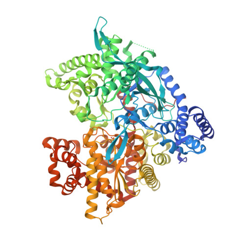
Crystallographic and computational studies on 4-phenyl-N-(beta-D-glucopyranosyl)-1H-1,2,3-triazole-1-acetamide, an inhibitor of glycogen phosphorylase: Comparison with alpha-D-glucose, N-acetyl-beta-D-glucopyranosylamine and N-benzoyl-N'-beta-D-glucopyranosyl urea binding.
Alexacou, K.M., Hayes, J.M., Tiraidis, C., Zographos, S.E., Leonidas, D.D., Chrysina, E.D., Archontis, G., Oikonomakos, N.G., Paul, J.V., Varghese, B., Loganathan, D.(2007) Proteins 71: 1307-1323
- PubMed: 18041758 Search on PubMed
- DOI: https://doi.org/10.1002/prot.21837
- Primary Citation Related Structures:
2PYD, 2PYI - PubMed Abstract:
4-Phenyl-N-(beta-D-glucopyranosyl)-1H-1,2,3-triazole-1-acetamide (glucosyltriazolylacetamide) has been studied in kinetic and crystallographic experiments with glycogen phosphorylase b (GPb), in an effort to utilize its potential as a lead for the design of potent antihyperglycaemic agents. Docking and molecular dynamics (MD) calculations have been used to monitor more closely the binding modes in operation and compare the results with experiment. Kinetic experiments in the direction of glycogen synthesis showed that glucosyltriazolylacetamide is a better inhibitor (K(i) = 0.18 mM) than the parent compound alpha-D-glucose (K(i) = 1.7 mM) or beta-D-glucose (K(i) = 7.4 mM) but less potent inhibitor than the lead compound N-acetyl-beta-D-glucopyranosylamine (K(i) = 32 microM). To elucidate the molecular basis underlying the inhibition of the newly identified compound, we determined the structure of GPb in complex with glucosyltriazolylacetamide at 100 K to 1.88 A resolution, and the structure of the compound in the free form. Glucosyltriazolylacetamide is accommodated in the catalytic site of the enzyme and the glucopyranose interacts in a manner similar to that observed in the GPb-alpha-D-glucose complex, while the substituent group in the beta-position of the C1 atom makes additional hydrogen bonding and van der Waals interactions to the protein. A bifurcated donor type hydrogen bonding involving O3H, N3, and N4 is seen as an important structural motif strengthening the binding of glucosyltriazolylacetamide with GP which necessitated change in the torsion about C8-N2 bond by about 62 degrees going from its free to the complex form with GPb. On binding to GP, glucosyltriazolylacetamide induces significant conformational changes in the vicinity of this site. Specifically, the 280s loop (residues 282-288) shifts 0.7 to 3.1 A (CA atoms) to accommodate glucosyltriazolylacetamide. These conformational changes do not lead to increased contacts between the inhibitor and the protein that would improve ligand binding compared with the lead compound. In the molecular modeling calculations, the GOLD docking runs with and without the crystallographic ordered cavity waters using the GoldScore scoring function, and without cavity waters using the ChemScore scoring function successfully reproduced the crystallographic binding conformation. However, the GLIDE docking calculations both with (GLIDE XP) and without (GLIDE SP and XP) the cavity water molecules were, impressively, further able to accurately reproduce the finer details of the GPb-glucosyltriazolylacetamide complex structure. The importance of cavity waters in flexible receptor MD calculations compared to "rigid" (docking) is analyzed and highlighted, while in the MD itself very little conformational flexibility of the glucosyltriazolylacetamide ligand was observed over the time scale of the simulations.
- Institute of Organic and Pharmaceutical Chemistry, National Hellenic Research Foundation, 116 35 Athens, Greece.
Organizational Affiliation: