2PFI

Crystal structure of the cytoplasmic domain of the human chloride channel ClC-Ka


Experimental Data Snapshot

  • Method: X-RAY DIFFRACTION
  • Resolution: 1.60 Å
  • R-Value Free: 0.218 
  • R-Value Work: 0.200 
  • R-Value Observed: 0.200 

wwPDB Validation   3D Report Full Report


This is version 1.3 of the entry. See complete history


Literature

The Structure of the Cytoplasmic Domain of the Chloride Channel ClC-Ka Reveals a Conserved Interaction Interface.

Markovic, S.Dutzler, R.

(2007) Structure 15: 715-725

  • DOI: https://doi.org/10.1016/j.str.2007.04.013
  • Primary Citation of Related Structures:  
    2PFI

  • PubMed Abstract: 

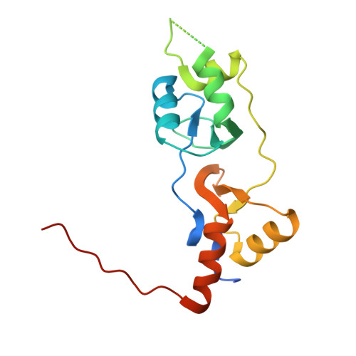
    The cytoplasmic domains of ClC chloride channels and transporters are ubiquitously found in eukaryotic family members and have been suggested to be involved in the regulation of ion transport. All cytoplasmic ClC domains share a conserved scaffold that contains a pair of CBS motifs. Here we describe the structure of the cytoplasmic component of the human chloride channel ClC-Ka at 1.6 A resolution. The structure reveals a dimeric organization of the domain that is unusual for CBS motif containing proteins. Using a biochemical approach combining mutagenesis, crosslinking, and analytical ultracentrifugation, we demonstrate that the interaction interface is preserved in solution and that the distantly related channel ClC-0 likely exhibits a similar structural organization. Our results reveal a conserved interaction interface that relates the cytoplasmic domains of ClC proteins and establish a structural relationship that is likely general for this important family of transport proteins.


  • Organizational Affiliation

    Department of Biochemistry, University of Zürich, Winterthurer Strasse 190, CH-8057 Zürich, Switzerland.


Macromolecules
Find similar proteins by:  (by identity cutoff)  |  3D Structure
Entity ID: 1
MoleculeChains Sequence LengthOrganismDetailsImage
Chloride channel protein ClC-Ka
A, B
164Homo sapiensMutation(s): 0 
Gene Names: CLCNKA
UniProt & NIH Common Fund Data Resources
Find proteins for P51800 (Homo sapiens)
Explore P51800 
Go to UniProtKB:  P51800
PHAROS:  P51800
GTEx:  ENSG00000186510 
Entity Groups  
Sequence Clusters30% Identity50% Identity70% Identity90% Identity95% Identity100% Identity
UniProt GroupP51800
Sequence Annotations
Expand
  • Reference Sequence
Experimental Data & Validation

Experimental Data

  • Method: X-RAY DIFFRACTION
  • Resolution: 1.60 Å
  • R-Value Free: 0.218 
  • R-Value Work: 0.200 
  • R-Value Observed: 0.200 
  • Space Group: P 1 21 1
Unit Cell:
Length ( Å )Angle ( ˚ )
a = 35.283α = 90
b = 83.421β = 92.48
c = 59.888γ = 90
Software Package:
Software NamePurpose
CNSrefinement
HKL-2000data collection
DENZOdata reduction
SCALEPACKdata scaling
SHELXSphasing

Structure Validation

View Full Validation Report


Entry History 

Deposition Data

Revision History  (Full details and data files)

  • Version 1.0: 2007-06-19
    Type: Initial release
  • Version 1.1: 2008-05-01
    Changes: Version format compliance
  • Version 1.2: 2011-07-13
    Changes: Version format compliance
  • Version 1.3: 2024-02-21
    Changes: Data collection, Database references, Derived calculations