Crystal structures and mechanism of 6-phospho-beta-galactosidase from Lactococcus lactis.
Wiesmann, C., Hengstenberg, W., Schulz, G.E.(1997) J Mol Biology 269: 851-860
- PubMed: 9223646 Search on PubMed
- DOI: https://doi.org/10.1006/jmbi.1997.1084
- Primary Citation Related Structures:
2PBG, 3PBG, 4PBG - PubMed Abstract:
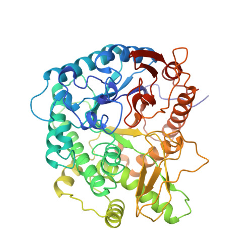
The initial structural model of 6-phospho-beta-galactosidase from Lactococcus lactis was refined to an R-factor of 16.4% (R[free] = 23.6%) to 2.3 A resolution (1 A = 0.1 nm), and the structures of three other crystal forms were solved by molecular replacement. The four structural models are essentially identical. The catalytic center of the enzyme is approximately at the mass center of the molecule and can only be reached through a 20 A long channel, which is observed with an "open" or "closed" entrance. The closed entrance is probably too small for the educt lactose-6-phosphate to enter, but large enough for the first product glucose to leave. Among the presented structures is a complex between an almost inactive mutant and the second product galactose-6-phosphate, which is exclusively bound at side-chains. A superposition (onto the native enzyme) of galactose-6-phosphate as bound to the mutant suggests the geometry of a postulated covalent intermediate. The binding mode of the educt was modeled, starting from the bound galactose-6-phosphate. A tightly fixed tryptophan is used as a chopping-board for splitting the disaccharide, and several other aromatic residues in the active center cavity are likely to participate in substrate transport/binding.
- Institut für Organische Chemie und Biochemie, Albert-Ludwigs-Universität, Freiburg im Breisgau, Germany.
Organizational Affiliation: