2P0B

Crystal structure of chemically-reduced E.coli nrfB


Experimental Data Snapshot

  • Method: X-RAY DIFFRACTION
  • Resolution: 1.74 Å
  • R-Value Free: 0.192 
  • R-Value Work: 0.159 
  • R-Value Observed: 0.161 

wwPDB Validation   3D Report Full Report


Ligand Structure Quality Assessment 


This is version 1.2 of the entry. See complete history


Literature

The crystal structure of the pentahaem c-type cytochrome NrfB and characterization of its solution-state interaction with the pentahaem nitrite reductase NrfA.

Clarke, T.A.Cole, J.A.Richardson, D.J.Hemmings, A.M.

(2007) Biochem J 406: 19-30

  • DOI: https://doi.org/10.1042/BJ20070321
  • Primary Citation of Related Structures:  
    2OZY, 2P0B

  • PubMed Abstract: 

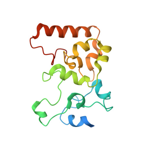
    NrfB is a small pentahaem electron-transfer protein widely involved in the respiratory reduction of nitrite or nitric oxide to ammonia, processes that provide energy for anaerobic metabolism in many enteric bacteria and also serve to detoxify these reactive nitrogen species. The X-ray crystal structure of Escherichia coli NrfB is presented at 1.74 A (1 A=0.1 nm) resolution. The architecture of the protein is that of a 40 A 'nanowire' in which the five haems are positioned within 6 A of each other along a polypeptide scaffold. During nitrite reduction, the physiological role of NrfB is to mediate electron transfer to another pentahaem protein, NrfA, the enzyme that catalyses periplasmic nitrite or nitric oxide reduction. Protein-protein interaction studies suggest NrfA and NrfB can form a 20-haem NrfA2-NrfB2 heterotetrameric complex.


  • Organizational Affiliation

    Centre for Metalloprotein Spectroscopy and Biology, School of Biological Sciences, University of East Anglia, Norwich NR4 7TJ, UK.


Macromolecules
Find similar proteins by:  (by identity cutoff)  |  3D Structure
Entity ID: 1
MoleculeChains Sequence LengthOrganismDetailsImage
Cytochrome c-type protein nrfB163Escherichia coli K-12Mutation(s): 0 
Gene Names: nrfB
UniProt
Find proteins for P0ABL1 (Escherichia coli (strain K12))
Explore P0ABL1 
Go to UniProtKB:  P0ABL1
Entity Groups  
Sequence Clusters30% Identity50% Identity70% Identity90% Identity95% Identity100% Identity
UniProt GroupP0ABL1
Sequence Annotations
Expand
  • Reference Sequence
Experimental Data & Validation

Experimental Data

  • Method: X-RAY DIFFRACTION
  • Resolution: 1.74 Å
  • R-Value Free: 0.192 
  • R-Value Work: 0.159 
  • R-Value Observed: 0.161 
  • Space Group: P 21 21 21
Unit Cell:
Length ( Å )Angle ( ˚ )
a = 49.727α = 90
b = 59.433β = 90
c = 65.318γ = 90
Software Package:
Software NamePurpose
SCALAdata scaling
REFMACrefinement
PDB_EXTRACTdata extraction
ADSCdata collection
MOSFLMdata reduction
MOLREPphasing

Structure Validation

View Full Validation Report


Ligand Structure Quality Assessment 


Entry History 

Deposition Data

Revision History  (Full details and data files)

  • Version 1.0: 2007-08-07
    Type: Initial release
  • Version 1.1: 2011-07-13
    Changes: Source and taxonomy, Version format compliance
  • Version 1.2: 2024-04-03
    Changes: Data collection, Database references, Derived calculations, Refinement description