Binding of sulfonium-ion analogues of di-epi-swainsonine and 8-epi-lentiginosine to Drosophila Golgi alpha-mannosidase II: The role of water in inhibitor binding.
Kumar, N.S., Kuntz, D.A., Wen, X., Pinto, B.M., Rose, D.R.(2008) Proteins 71: 1484-1496
- PubMed: 18076078 Search on PubMed
- DOI: https://doi.org/10.1002/prot.21850
- Primary Citation Related Structures:
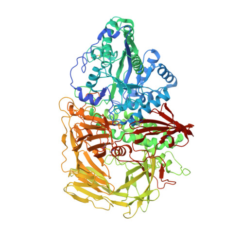
2OW6, 2OW7 - PubMed Abstract:
Retaining glycosidases operate by a two-step catalytic mechanism in which the transition states are characterized by buildup of a partial positive charge at the anomeric center. Sulfonium-ion analogues of the naturally occurring glycosidase inhibitors, swainsonine and 8-epi-lentiginosine, in which the bridgehead nitrogen atom is replaced by a sulfonium-ion, were synthesized in order to test the hypothesis that a sulfonium salt carrying a permanent positive charge would be an effective glycosidase inhibitor. Initial prediction based on computational docking indicated three plausible binding modes to Drosophila Golgi alpha-mannosidase II (dGMII), the most likely being close to that of swainsonine. Observation of the binding of di-epi-thioswainsonine and 8-epi-thiolentiginosine to dGMII from crystallographic data, however, revealed an orientation different from swainsonine in the active site. Screening these two compounds against dGMII shows that they are inhibitors with IC(50) values of 2.0 and 0.014 mM, respectively. This dramatic difference in affinity between the two compounds, which differ by only one hydroxyl group, is rationalized in terms of bound water molecules and the water molecule substructure in the active site, as identified by comparison of high resolution X-ray crystal structures of several dGMII-inhibitor complexes.
- Department of Chemistry, Simon Fraser University, Burnaby, British Columbia, Canada V5A 1S6.
Organizational Affiliation: